- Respiratory system
-
See also: Respiratory tract
Respiratory 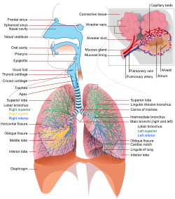
A complete, schematic view of the human respiratory system with their parts and functions. Latin systema respiratorium The respiratory system is the anatomical system of an organism that introduces respiratory gases to the interior and performs gas exchange. In humans and other mammals, the anatomical features of the respiratory system include airways, lungs, and the respiratory muscles. Molecules of oxygen and carbon dioxide are passively exchanged, by diffusion, between the gaseous external environment and the blood. This exchange process occurs in the alveolar region of the lungs.[1] Other animals, such as insects, have respiratory systems with very simple anatomical features, and in amphibians even the skin plays a vital role in gas exchange. Plants also have respiratory systems but the directionality of gas exchange can be opposite to that in animals. The respiratory system in plants also includes anatomical features such as holes on the undersides of leaves known as stomata.[2]
Contents
Comparative anatomy and physiology
Horses
Horses are obligate nasal breathers which means that they are different from many other mammals because they do not have the option of breathing through their mouths and must take in oxygen through their noses.
Elephants
The elephant is the only animal known to have no pleural space. Rather, the parietal and visceral pleura are both composed of dense connective tissue and joined to each other via loose connective tissue.[3] This lack of a pleural space, along with an unusually thick diaphragm, are thought to be evolutionary adaptations allowing the elephant to remain underwater for long periods of time while breathing through its trunk which emerges as a snorkel.[4]
Birds
The respiratory system of birds differs significantly from that found in mammals, containing unique anatomical features such as air sacs. The lungs of birds also do not have the capacity to inflate as birds lack a diaphragm and a pleural cavity. Gas exchange in birds occurs between air capillaries and blood capillaries, rather than in alveoli. See Avian respiratory system for a detailed description of these and other features.
Reptiles
The anatomical structure of the lungs is less complex in reptiles than in mammals, with reptiles lacking the very extensive airway tree structure found in mammalian lungs. Gas exchange in reptiles still occurs in alveoli however, reptiles do not possess a diaphragm. Thus, breathing occurs via a change in the volume of the body cavity which is controlled by contraction of intercostal muscles in all reptiles except turtles. In turtles, contraction of specific pairs of flank muscles governs inspiration or expiration.[5]
See also reptiles for more detailed descriptions of the respiratory system in these animals.
Amphibians
Both the lungs and the skin serve as respiratory organs in amphibians. The skin of these animals is highly vascularized and moist, with moisture maintained via secretion of mucus from specialized cells. While the lungs are of primary importance to breathing control, the skin's unique properties aid rapid gas exchange when amphibians are submerged in oxygen-rich water.[6]
Fish
In most fish respiration takes place through gills. (See also aquatic respiration.) Lungfish, however, do possess one or two lungs. The labyrinth fish have developed a special organ that allows them to take advantage of the oxygen of the air.
Anatomy in invertebrates
Insects
Air enters the respiratory systems of most insects through a series of external openings called spiracles. These external openings, which act as muscular valves in some insects, lead to the internal respiratory system, a densely networked array of tubes called tracheae. The scientific tracheal system within an individual is composed of interconnecting transverse and longitudinal tracheae which maintain equivalent pressure throughout the system. These tracheae branch repeatedly, eventually forming tracheoles, which are blind-ended, water-filled compartments only one micrometer in diameter.[7] It is at this level of the tracheoles that oxygen is delivered to the cells for respiration. The trachea are water-filled due to the permeable membrane of the surrounding tissues. During exercise, the water level retracts due to the increase in concentration of lactic acid in the muscle cells. This lowers the water potential and the water is drawn back into the cells via osmosis and air is brought closer to the muscle cells. The diffusion pathway is then reduced and gases can be transferred more easily.
Insects were once believed to exchange gases with the environment continuously by the simple diffusion of gases into the tracheal system. More recently, however, large variation in insect ventilatory patterns have been documented and insect respiration appears to be highly variable. Some small insects do demonstrate continuous respiration and may lack muscular control of the spiracles. Others, however, utilize muscular contraction of the abdomen along with coordinated spiracle contraction and relaxation to generate cyclical gas exchange patterns and to reduce water loss into the atmosphere. The most extreme form of these patterns is termed discontinuous gas exchange cycles (DGC).[8]
Mollusks
Mollusks generally possess gills that allow exchange of oxygen from an aqueous environment into the circulatory system. These animals also possess a heart that pumps blood which contains hemocyaninine as its oxygen-capturing molecule. Hence, this respiratory system is similar to that of vertebrate fish. The respiratory system of gastropods can include either gills or a lung.
Physiology in mammals
For more detailed descriptions see also Respiratory physiology or Respiration.
Ventilation
In respiratory physiology, ventilation (or ventilation rate) is the rate at which gas enters or leaves the lung. It is categorised under the following definitions:
Measurement Equation Description Minute ventilation tidal volume * respiratory rate[1][2] the total volume of gas entering the lungs per minute. Alveolar ventilation (tidal volume - dead space) * respiratory rate [1] the volume of gas per unit time that reaches the alveoli, the respiratory portions of the lungs where gas exchange occurs. Dead space ventilation dead space * respiratory rate[3] the volume of gas per unit time that does not reach these respiratory portions, but instead remains in the airways (trachea, bronchi, etc.). Control
Ventilation occurs under the control of the autonomic nervous system from parts of the brain stem, the medulla oblongata and the pons. This area of the brain forms the respiration regulatory center, a series of interconnected brain cells within the lower and middle brain stem which coordinate respiratory movements. The sections are the pneumotaxic center, the apneustic center, and the dorsal and ventral respiratory groups. This section is especially sensitive during infancy, and the neurons can be destroyed if the infant is dropped and/or shaken violently. The result can be death due to "shaken baby syndrome".[9]
Inhalation
Inhalation is initiated by the diaphragm and supported by the external intercostal muscles. Normal resting respirations are 10 to 18 breaths per minute, with a time period of 2 seconds. During vigorous inhalation (at rates exceeding 35 breaths per minute), or in approaching respiratory failure, accessory muscles of respiration are recruited for support. These consist of sternocleidomastoid, platysma, and the scalene muscles of the neck. Pectoral muscles and latissimus dorsi are also accessory muscles.
Under normal conditions, the diaphragm is the primary driver of inhalation. When the diaphragm contracts, the ribcage expands and the contents of the abdomen are moved downward. This results in a larger thoracic volume and negative pressure (with respect to atmospheric pressure) inside the thorax. As the pressure in the chest falls, air moves into the conducting zone. Here, the air is filtered, warmed, and humidified as it flows to the lungs.
During forced inhalation, as when taking a deep breath, the external intercostal muscles and accessory muscles aid in further expanding the thoracic cavity. During inhalation the diaphragm contracts.
Exhalation
Exhalation is generally a passive process; however, active or forced exhalation is achieved by the abdominal and the internal intercostal muscles. During this process air is forced or exhaled out.
The lungs have a natural elasticity: as they recoil from the stretch of inhalation, air flows back out until the pressures in the chest and the atmosphere reach equilibrium.[10]
During forced exhalation, as when blowing out a candle, expiratory muscles including the abdominal muscles and internal intercostal muscles, generate abdominal and thoracic pressure, which forces air out of the lungs.
Gas exchange
The major function of the respiratory system is gas exchange between the external environment and an organism's circulatory system. In humans and mammals, this exchange facilitates oxygenation of the blood with a concomitant removal of carbon dioxide and other gaseous metabolic wastes from the circulation. As gas exchange occurs, the acid-base balance of the body is maintained as part of homeostasis. If proper ventilation is not maintained, two opposing conditions could occur: respiratory acidosis, a life threatening condition, and respiratory alkalosis.
Upon inhalation, gas exchange occurs at the alveoli, the tiny sacs which are the basic functional component of the lungs. The alveolar walls are extremely thin (approx. 0.2 micrometres). These walls are composed of a single layer of epithelial cells (type I and type II epithelial cells) close to the pulmonary capillaries which are composed of a single layer of endothelial cells. The close proximity of these two cell types allows permeability to gases and, hence, gas exchange. This whole mechanism of gas exchange is carried by the simple phenomenon of pressure difference. When the atmospheric pressure is low outside, the air from lungs flow out. When the air pressure is low inside, then the vice versa.
Non-respiratory functions
Lung Defense Mechanisms
Airway epithelial cells can secrete a variety of molecules that aid in lung defense. Secretory immunoglobulins (IgA), collectins (including Surfactant A and D), defensins and other peptides and proteases, reactive oxygen species, and reactive nitrogen species are all generated by airway epithelial cells. These secretions can act directly as antimicrobials to help keep the airway free of infection. Airway epithelial cells also secrete a variety of chemokines and cytokines that recruit the traditional immune cells and others to site of infections.
Metabolic & Endocrine Functions of the Lungs
In addition to their functions in gas exchange, the lungs have a number of metabolic functions. They manufacture surfactant for local use, as noted above. They also contain a fibrinolytic system that lyses clots in the pulmonary vessels. They release a variety of substances that enter the systemic arterial blood and they remove other substances from the systemic venous blood that reach them via the pulmonary artery. Prostaglandins are removed from the circulation, but they are also synthesized in the lungs and released into the blood when lung tissue is stretched. The lungs also activate one hormone; the physiologically inactive decapeptide angiotensin I is converted to the pressor, aldosterone-stimulating octapeptide angiotensin II in the pulmonary circulation. The reaction occurs in other tissues as well, but it is particularly prominent in the lungs. Large amounts of the angiotensin-converting enzyme responsible for this activation are located on the surface of the endothelial cells of the pulmonary capillaries. The converting enzyme also inactivates bradykinin. Circulation time through the pulmonary capillaries is less than 1 s, yet 70% of the angiotensin I reaching the lungs is converted to angiotensin II in a single trip through the capillaries. Four other peptidases have been identified on the surface of the pulmonary endothelial cells.
Vocalization
The movement of gas through the larynx, pharynx and mouth allows humans to speak, or phonate. Vocalization, or singing, in birds occurs via the syrinx, an organ located at the base of the trachea. The vibration of air flowing across the larynx (vocal chords), in humans, and the syrinx, in birds, results in sound. Because of this, gas movement is extremely vital for communication purposes.
Temperature control
Panting in dogs and some other animals provides a means of controlling body temperature. This physiological response is used as a cooling mechanism.
Coughing and sneezing
Irritation of nerves within the nasal passages or airways, can induce coughing and sneezing. These responses cause air to be expelled forcefully from the trachea or nose, respectively. In this manner, irritants caught in the mucus which lines the respiratory tract are expelled or moved to the mouth where they can be swallowed.
Development in People
Humans and mammals
Further information: Development of human lungThe respiratory system lies dormant in the human fetus during pregnancy. At birth, the respiratory system becomes fully functional upon exposure to air, although some lung development and growth continues throughout childhood. Pre-term birth can lead to infants with under-developed lungs. These lungs show incomplete development of the alveolar type II cells, cells that produce surfactant. The lungs of pre-term infants may not function well because the lack of surfactant leads to increased surface tension within the alveoli. Thus, many alveoli collapse such that no gas exchange can occur within some or most regions of an infant's lungs, a condition termed respiratory distress syndrome. Basic scientific experiments, carried out using cells from chicken lungs, support the potential for using steroids as a means of furthering development of type II alveolar cells.[11] In fact, once a pre-mature birth is threatened, every effort is made to delay the birth, and a series of steroid shots is frequently administered to the mother during this delay in an effort to promote lung growth.[12]
Disease
Disorders of the respiratory system can be classified into four general areas:
- Obstructive conditions (e.g., emphysema, bronchitis, asthma)
- Restrictive conditions (e.g., fibrosis, sarcoidosis, alveolar damage, pleural effusion)
- Vascular diseases (e.g., pulmonary edema, pulmonary embolism, pulmonary hypertension)
- Infectious, environmental and other "diseases" (e.g., pneumonia, tuberculosis, asbestosis, particulate pollutants):
Coughing is of major importance, as it is the body's main method to remove dust, mucus, saliva, and other debris from the lungs. Inability to cough can lead to infection. Deep breathing exercises may help keep finer structures of the lungs clear from particulate matter, etc.
The respiratory tract is constantly exposed to microbes due to the extensive surface area, which is why the respiratory system includes many mechanisms to defend itself and prevent pathogens from entering the body.
Disorders of the respiratory system are usually treated internally by a pulmonologist and Respiratory Therapist.
Plants
Plants use carbon dioxide gas in the process of photosynthesis, and exhale oxygen gas as waste. The chemical equation of photosynthesis is 6 CO2 (carbon dioxide) and 6 H2O (water) and that makes 6 O2 (oxygen) and C6H12O6 (glucose). Respiration is the opposite of that. However, plants also sometimes respire as humans do, taking in oxygen and producing carbon dioxide.
Plant respiration is limited by the process of diffusion. Plants take in carbon dioxide through holes on the undersides of their leaves known as stoma or pores. However, most plants require little air.[citation needed] Most plants have relatively few living cells outside of their surface because air (which is required for metabolic content) can penetrate only skin deep. However, most plants are not involved in highly aerobic activities, and thus have no need of these living cells.
Teamwork
Circulatory System
obviously it interacts with the circulatory system because the lungs are where the oxygen is picked up by the blood and then transported around the body hemoglobin molecules in the red blood cells pick up oxygen where it is abundant (the lungs) and deliver it to respiring tissues (muscles...) so it also interacts with the muscle system
Nervous System
it also interacts with the nervous system because this is what controls the breathing rate, the breathing rate needs to be changed when there is too high concentration of carbon dioxide. when the concentration of CO2 increases the chemo-receptor (chemical sensitive) cells in the wall of the carotid artery and aorta sends impulses to the respiratory center of the brain, nerve impulses are also sent to the respiratory center from the stretch receptors in the lungs - the more the lungs inflate the more nerve impulses are sent to the respiratory center when the respiratory center receives these impulses it sends impulses to the diaphragm and intercostal muscles causing them to contract and making the breathing rate increase
Immune System
Most of the respiratory system is lined with mucous membranes which contain mucosal-associated lymphoid tissue, this tissue is part of the lymphatic system which is an essential part of the immune system because it produces immune cells (e.g. Lymphocyte which is a type of white blood cell) lymphocytes just defend the body against infections and viruses.
References
- ^ Haton, Anthea; Jean, Hopkins Susan, Johnson Charles William, McLaughlin Maryanna Quon Warner David, LaHart Wright, Jill D. (2009). Human Biology and Health. Englewood Cliffs,: Prentice Hall. pp. 108–118. ISBN 0-12-981176-1.
- ^ West, John B.. Respiratory physiology-- the essentials. Baltimore: Williams & Wilkins. pp. 1–10. ISBN 0-683-08937-4.
- ^ West, John B.; Ravichandran (1993). "Snorkel breathing in the elephant explains the unique anatomy of its pleura". Respiration Physiology 126 (1): 1–8. doi:10.1016/S0034-5687(01)00203-1. PMID 11311306.
- ^ West, John B. (2002). "Why doesn't the elephant have a pleural space?". News Physiol Sci 17: 47–50. PMID 11909991.
- ^ Britannica On-line Encyclopedia
- ^ Gottlieb, G; Jackson DC (1976). "Importance of pulmonary ventilation in respiratory control in the bullfrog". Am J Physiol 230 (3): 608–13. PMID 4976.
- ^ Introduction to Insect Anatomy
- ^ Lighton, JRB (January 1996). "Discontinuous gas exchange in insects". Annu Rev Entomology 41: 309–324.
- ^ *Fact sheet on Shaken Baby Syndrome
- ^ A simple model of how the lungs are inflated can be built from a bell jar
- ^ Department of Environmental Biology, University of Adelaide, Adelaide, South Australia
- ^ Pregnancy-facts.com
External links
- A high school level description of the respiratory system
- Introduction to Respiratory System
- Science aid: Respiratory System A simple guide for high school students
- The Respiratory System University level (Microsoft Word document)
Human systems and organs TA 2–4:
MSBone (Carpus · Collar bone (clavicle) · Thigh bone (femur) · Fibula · Humerus · Mandible · Metacarpus · Metatarsus · Ossicles · Patella · Phalanges · Radius · Skull (cranium) · Tarsus · Tibia · Ulna · Rib · Vertebra · Pelvis · Sternum) · CartilageTA 5–11:
splanchnic/
viscusmostly
ThoracicRespiratory systemmostly
AbdominopelvicDigestive system+
adnexaMouth (Salivary gland, Tongue) · upper GI (Oropharynx, Laryngopharynx, Esophagus, Stomach) · lower GI (Small intestine, Appendix, Colon, Rectum, Anus) · accessory (Liver, Biliary tract, Pancreas)TA 12–16 Blood
(Non-TA)General anatomy: systems and organs, regional anatomy, planes and lines, superficial axial anatomy, superficial anatomy of limbsHead and neck, upper RT: Nose (TA A06.1, TH H3.05.01, GA 10.992) External nose Ala of nose
nasal cartilages (of the septum, Greater alar, Lesser alar, Lateral nasal, Accessory nasal, Vomeronasal)Nasal cavity OpeningsLateral wallNasal concha/meati: Superior nasal concha · Middle nasal concha · Inferior nasal concha · Superior nasal meatus · Middle nasal meatus · Inferior nasal meatus
Sphenoethmoidal recess · Ethmoid bulla · Agger nasi · Ethmoidal infundibulum · Semilunar hiatus · Maxillary hiatusMedial wallParanasal sinuses Naso-pharynx Head and neck anatomy, Upper RT: Larynx (TA A06.2, TH H3.05.01, GA 11.1072) Cartilages major/unpaired: Epiglottis (Vallecula) · Thyroid (Laryngeal prominence, Oblique line, Superior thyroid notch, Superior horn, Inferior horn) · Cricoid
minor/paired: Arytenoid (Vocal process, Muscular process) · Corniculate · CuneiformLigaments/folds extrinsic ligaments: Hyoepiglottic ligament · Thyrohyoid membrane (Lateral ligament, Median ligament) · Thyroepiglottic ligament · Cricotracheal ligament
intrinsic ligaments · upper: Quadrangular membrane (Aryepiglottic, Vestibular ligament/Vestibular fold)
intrinsic ligaments · lower: Cricothyroid ligament (Median, Lateral/Conus elasticus, Vocal ligament/Vocal folds)Laryngeal cavity Other Anatomy: Lower RT respiratory system (TA A06.3–5, TH H3.05.02, GA 11.1084) TB tree main bronchus (right, left) · lobar/secondary bronchi (eparterial bronchus) · segmental/tertiary bronchiLungs GeneralLeft lung/Right lung · Base/Apex · Root/Hilum
Superior lobe · Lingula of left lung/Middle lobe of right lung · Inferior lobe
borders: Anterior border (Cardiac notch) · Posterior border · Inferior border
surfaces: Costal surface · Mediastinal surface (Cardiac impression) · Diaphragmatic surface
fissures: Oblique fissure · Horizontal fissureBronchiole: Conducting zone (Terminal bronchiole) · Respiratory zone (Respiratory bronchiole · Alveolar duct · Alveolus · Blood-air barrier)CellsAnatomy, Respiratory system: Thoracic cavity (TA A07, TH H3.05.03, GA 11.1087) Pleurae Mediastinum General Pathology of respiratory system (J, 460–519), respiratory diseases Upper RT
(including URTIs,
Common cold)HeadLower RT/lung disease
(including LRTIs)acute: Acute bronchitischronic: COPD (Chronic bronchitis, Acute exacerbations of chronic bronchitis, Acute exacerbation of COPD, Emphysema) · Asthma (Status asthmaticus, Aspirin-induced, Exercise-induced) · BronchiectasisPneumoconiosis (Asbestosis, Baritosis, Bauxite fibrosis, Berylliosis, Caplan's syndrome, Chalicosis, Coalworker's pneumoconiosis, Siderosis, Silicosis, Talcosis, Byssinosis)
Hypersensitivity pneumonitis (Bagassosis, Bird fancier's lung, Farmer's lung, Lycoperdonosis)OtherARDS · Pulmonary edema · Löffler's syndrome/Eosinophilic pneumonia · Respiratory hypersensitivity (Allergic bronchopulmonary aspergillosis)
Hamman-Rich syndrome · Idiopathic pulmonary fibrosis · SarcoidosisObstructive or
restrictiveBy pathogenViral · Bacterial (Pneumococcal, Klebsiella) / Atypical bacterial (Mycoplasma, Legionnaires' disease, Chlamydiae) · Fungal (Pneumocystis) · Parasitic · noninfectious (Chemical/Mendelson's syndrome, Aspiration/Lipid)By vector/routeBy distributionBroncho- · LobarOtherPleural cavity/
mediastinumMediastinal diseaseOther/general Prenatal development/Mammalian development of respiratory system (overview) (GA 11.1071, TE E5.5) Upper Lower Laryngotracheal groove · Respiratory budCategories:

Wikimedia Foundation. 2010.