- Sarcoidosis
-
Sarcoidosis Classification and external resources 
Chest xray showing the typical nodularity of sarcoidosis in the base of the lungs.ICD-10 D86 ICD-9 135 OMIM 181000 DiseasesDB 11797 MedlinePlus 000076 eMedicine med/2063 MeSH D012507 Sarcoidosis (from sarc meaning flesh, -oid, like, and -osis, diseased or abnormal condition), also called sarcoid, Besnier-Boeck disease or Besnier-Boeck-Schaumann disease, is a disease in which abnormal collections of chronic inflammatory cells (granulomas) form as nodules in multiple organs.[1] The cause of sarcoidosis is unknown. Granulomas most often appear in the lungs or the lymph nodes, but virtually any organ can be affected. Normally the onset is gradual. Sarcoidosis may be asymptomatic or chronic. It commonly improves or clears up spontaneously. More than 2/3 of people with lung sarcoidosis have no symptoms after 9 years. About 50% have relapses. About 10% develop serious disability. Lung scarring or infection may lead to respiratory failure and death.[1] Chronic patients may deal with waxing and waning symptoms over many years.[2]
Contents
Signs and symptoms
 Signs and symptoms of sarcoidosis.[3]
 Systemic sarcoidosis
Sarcoidosis is a systemic disease that can affect any organ. Common symptoms are vague, such as fatigue unchanged by sleep, lack of energy, weight loss, aches and pains, arthritis, dry eyes, swelling of the knees, blurry vision, shortness of breath, a dry hacking cough or skin lesions. Sarcoidosis and cancer may mimic one another, making the distinction difficult.[4] The cutaneous symptoms vary, and range from rashes and noduli (small bumps) to erythema nodosum or lupus pernio. It is often asymptomatic.
The combination of erythema nodosum, bilateral hilar lymphadenopathy and arthralgia is called Löfgren syndrome. This syndrome has a relatively good prognosis.
Renal, liver (including portal hypertension), heart[5] or brain involvement may cause further symptoms and altered functioning.
Lungs
Of individuals with sarcoidosis, 90 percent have an abnormal chest x-ray at some time during their course. Overall, approximately 50 percent develop permanent pulmonary abnormalities and 5 to 15 percent have progressive fibrosis of the lung parenchyma. Sarcoidosis of the lung is primarily an interstitial lung disease in which the inflammatory process involves the aveoli, small bronchi, and small blood vessels. In acute and subacute cases, the physical examination usually reveals dry rales.[6]
Liver
Although liver biopsy reveals liver involvement in 60 to 90 percent of cases, liver dysfunction is usually not important clinically. Approximately 20-30% have hepatomegaly and/or biochemical evidence of liver involvement. Usually these changes reflect a cholestatic pattern and include an elevated alkaline phosphatase level; the bilirubin and aminotransferases are only mildly elevated. Jaundice is rare.[6]
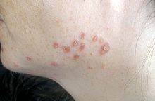
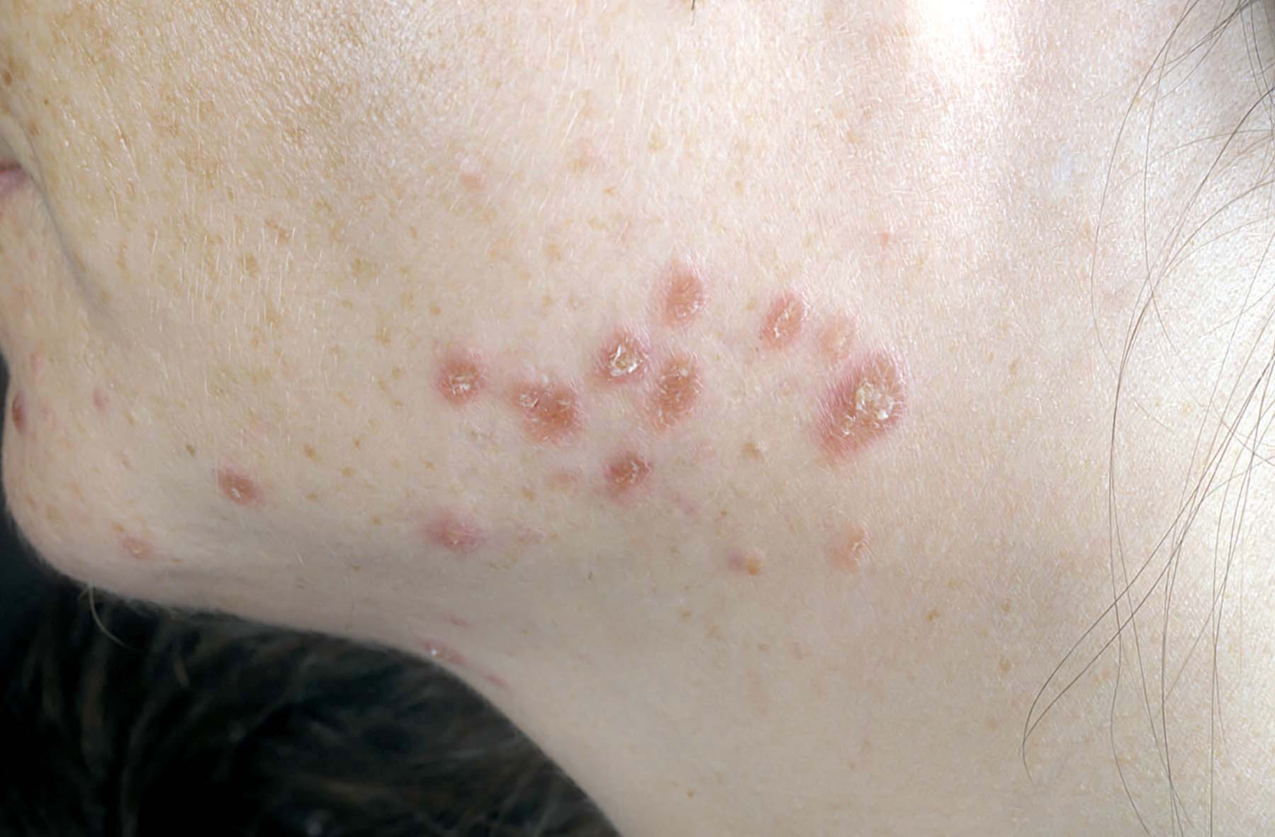
Skin
Sarcoidosis involves the skin in about 25 percent of patients. The most common lesions are erythema nodosum, plaques, maculopapular eruptions, subcutaneous nodules, and lupus pernio. Treatment is not required, since the lesions usually resolve spontaneously in 2 to 4 weeks. Although it may be disfiguring, cutaneous sarcoidosis rarely causes major problems.[6]
Heart
Although cardiac involvement is present in 20% to 30% of patients with sarcoidosis, only about 5% of patients with systemic sarcoidosis are symptomatic.[7]
The presentation of cardiac sarcoidosis can range from asymptomatic conduction abnormalities to fatal ventricular arrhythmia.[8] Myocardial sarcoidosis can be a rare cause of sudden cardiac death.[9][10]
Eye
Manifestations in the eye include uveitis, uveoparotitis, and retinal inflammation, which may result in loss of visual acuity or blindness. The combination of anterior uveitis, parotitis, VII cranial nerve paralysis and fever is called uveoparotid fever, and is associated with Heerfordt-Waldenstrom syndrome. (D86.8)
Blood
Abnormal clinical blood tests are frequent but not diagnostic. Anemia occurs in 4-20% of patients with sarcoidosis. Leukopenia (due to a reduced number of circulating lymphocytes [11] or lymphopenia) occurs in as many as 40% of patients but is rarely severe. In the absence of splenomegaly, leukopenia may reflect bone marrow involvement, however, the most common mechanism is a redistribution of blood T cells to sites of disease.[12] Other non-specific findings include monocytosis, occurring in the majority of sarcoidosis cases [13], increased hepatic enzymes or alkaline phosphatase. Hypercalciuria and hypercalcemia are seen in <10% of patients.[14]
Lymph nodes
Lymphadenopathy is very common in sarcoidosis. Intrathoracic nodes are enlarged in 75 to 90 percent of all patients; usually this involves the hilar nodes, but the paratracheal nodes are commonly involved. Peripheral lymphadenopathy is very common, particularly involving the cervical (the most common head and neck manifestation of the disease [15]), axillary, epitrochlear, and inguinal nodes. Palpation causes no pain.[6]
Nervous system
All components of the nervous system can be involved in sarcoidosis. Sarcoidosis affecting the brain or nerves is known as neurosarcoidosis. Neurologic findings are observed in about 5 percent of patients. Seventh nerve involvement with unilateral facial paralysis is most common. It occurs suddenly and is usually transient. Other common manifestations of neurosarcoid include optic nerve dysfunction, papilledema, palate dysfunction, hearing abnormalities, hypothalamic and pituitary abnormalities, chronic meningitis, and peripheral neuropathy [6]. Intramedullary sarcoidosis is rare and occurs in less than 1% of cases. There is usually granulomatous involvement of the basal meninges that subsequently affects the cranial nerves. Myelopathy may be the initial clinical presentation of intramedullary neurosarcoidosis.[16]
Exocrine glands
Parotid enlargement is a classic feature of sarcoidosis, but clinically apparent parotid involvement occurs in less than 10 percent of patients. Bilateral involvement is the rule. The gland is usually nontender, firm, and smooth. Xerostomia can occur; other exocrine glands are affected only rarely.[6]
Scalp
Sarcoidosis of the scalp presents with diffuse or patchy hair loss.[17]:762
Causes
The exact cause of sarcoidosis is not known. The current working hypothesis is that in genetically susceptible individuals sarcoidosis is caused through alteration in immune response after exposure to an environmental, occupational, or infectious agent.[18]
Genetics
Investigations of genetic susceptibility yielded many candidate genes but only few were confirmed by further investigations and no reliable genetic markers are known. Currently, the most interesting candidate gene is BTNL2; several HLA-DR risk alleles are also being investigated.[19] In persistent sarcoidosis the HLA haplotype HLA-B7-DR15 are either cooperating in disease or another gene between these two loci is associated. In non-persistent disease there is a strong genetic association with HLA DR3-DQ2.[20] Siblings have only a modestly increased risk (hazard ratio 5-6) of developing the disease, indicating that genetic susceptibility plays only a small role. The alternate hypothesis that family members share similar exposures to environmental pathogens is quite plausible to explain the apparent hereditary factor.
Infectious agents
Several infectious agents appear to be significantly associated with sarcoidosis but none of the known associations is specific enough to suggest a direct causative role. Propionibacterium acnes can be found in bronchoalveolar lavage of approximately 70% patients and is associated with disease activity, however it can be also found in 23% of controls.[21][22] A recent meta-analysis investigating the role of mycobacteria in sarcoidosis found it was present in 26.4% of cases, however the meta-analysis also detected a possible publication bias, so the results need further confirmation.[23][24]
There have also been reports of transmission of sarcoidosis via organ transplants.[25]
Vitamin D dysregulation
Sarcoidosis frequently causes an increase in vitamin D production outside the kidney.[26] Macrophages inside the granulomas convert vitamin D to its active form, resulting in elevated levels of the hormone 1,25-dihydroxyvitamin D and symptoms of hypervitaminosis D that may include fatigue, lack of strength or energy, irritability, metallic taste, temporary memory loss or cognitive problems. Physiological compensatory responses (e.g., suppression of the parathyroid hormone levels) may mean the patient does not develop frank hypercalcemia. This condition may be aggravated by high levels of estradiol and prolactin such as in pregnancy, leading to hypercalciuria and/or compensatory hypoparathyroidism.[27] High levels of Vitamin D are also implicated in immune-system dysfunctions which tie into the sarcoid condition.
Hyperprolactinemia
Prolactin is frequently increased in sarcoidosis, between 3–32% cases have hyperprolactinemia,[28] this frequently leads to amenorrhea, galactorrhea or nonpuerperal mastitis in women. Prolactin also has a broad spectrum of effects on the immune system and increased prolactin levels are associated with disease activity or may exacerbate symptoms in many autoimmune diseases and treatment with prolactin lowering medication has been shown effective in some cases.[29] However it is unknown if this relation holds in sarcoidosis and the gender predilection in sarcoidosis is less pronounced than in some other autoimmune diseases where such relation has been established. In pregnancy, the effects of prolactin and estrogen counteract each other to some degree, with a slight trend to improve pulmonary manifestations of sarcoidosis while lupus, uveitis and arthralgia might slightly worsen.[27] Lupus, uveitis and arthralgia are known to be in some cases associated with increased prolactin levels and respond to bromocriptin treatment but so far this has not been investigated specifically for sarcoidosis. The reasons for increased prolactin levels in sarcoidosis are uncertain. It has been observed that prolactin is produced by T-lymphocytes in some autoimmune disorders in amounts high enough to affect the feedback by the hypothalamic dopaminergic system.[30]
The extrapituitary prolactin is believed to play a role as a cytokine like proinflammatory factor. Prolactin antibodies are believed to play a role in hyperprolactinemia in other autoimmune disorders and high prevalence endocrine autoimmunity has been observed in patients with sarcoidosis.[31] It may also be a consequence of renal disease or treatment with steroids. Neurosarcoidosis may occasionally cause hypopituiarism but has not been reported to cause hyperprolactinemia.
Thyroid disease
In women, a substantial association of thyroid disease and sarcoidosis has been reported. The association is less marked but still significant for male patients. Female patients have a significantly elevated risk for hypothyroidism, hyperthyroidism and thyroid autoimmunity and it appears that autoimmunity is very important in the pathogenesis of thyroid disease in this population. Thyroid granulomatosis on the other hand is uncommon.[32]
Autoimmune
Association of autoimmune disorders has been frequently observed. The exact mechanism of this relation is not known but some evidence supports the hypothesis that this is a consequence of Th1 lymphokine prevalence.[33][32]
Sarcoidosis has been associated with celiac disease. Celiac disease is a condition in which there is a chronic reaction to certain protein chains, commonly referred to as glutens, found in some cereal grains. This reaction causes destruction of the villi in the small intestine, with resulting malabsorption of nutrients.
An association with type IV hypersensitivity has been described.[34] Tests of delayed cutaneous hypersensitivity have been used to measure progression.[35]
Other
While disputed, some cases have been associated with inhalation of the dust from the collapse of the World Trade Center after the September 11, 2001 attacks.[36] See Health effects arising from the September 11, 2001 attacks for more information. Chicago comedian, Bernie Mac, suffered from sarcoidosis and died of pneumonia as a result of his compromised immune system.[37] Reggie White, a former standout National Football League player, also suffered from sarcoidosis, and the disease played a major role in his death.[38]
Pathophysiology
Granulomatous inflammation is characterized primarily by accumulation of monocytes, macrophages and activated T-lymphocytes, with increased production of key inflammatory mediators, TNF-alpha, IFN-gamma, and IL-12, characteristic of a Th1-polarized response (T-helper lymphocyte-1 response). Sarcoidosis has paradoxical effects on inflammatory processes; it is characterized by increased macrophage and CD4 helper T-cell activation resulting in accelerated inflammation, however, immune response to antigen challenges such as tuberculin is suppressed. This paradoxic state of simultaneous hyper- and hypo- activity is suggestive of a state of anergy. The anergy may also be responsible for the increased risk of infections and cancer. It appears that regulatory T-lymphocytes in the periphery of sarcoid granulomas suppress IL-2 secretion which is hypothesized to cause the state of anergy by preventing antigen-specific memory responses.[39]
While it is widely believed that TNF-alpha plays an important role in the formation of granulomas, it was observed that sarcoidosis can be triggered by treatment with the TNF-alpha antagonist etanercept.[40][41]
-

Sarcoidosis in a lymph node.
-

Asteroid Body in sarcoidosis.
-

Micrograph showing pulmonary sarcoidosis with granulomas with asteroid bodies. H&E stain.
Diagnosis
Diagnosis of sarcoidosis is often a matter of exclusion. To exclude sarcoidosis in a case presenting with pulmonary symptoms might involve chest X-ray, CT scan of chest, PET scan, CT-guided biopsy, mediastinoscopy, open lung biopsy, bronchoscopy with biopsy, endobronchial ultrasound and endoscopic ultrasound with FNA of mediastinal lymph nodes(EBUS FNA). Tissue from biopsy of lymph nodes is subjected to both flow cytometry to rule out cancer and special stains (acid fast bacilli stain and Gömöri methenamine silver stain) to rule out microorganisms and fungi. Angiotensin-converting enzyme blood levels are used in diagnosis and monitoring of sarcoidosis.[42]
Differential diagnosis includes metastatic disease, lymphoma, septic emboli, rheumatoid nodules, Wegener's granulomatosis, varicella infection, and atypical infections such as mycobacterium avium complex, cytomegalovirus, and cryptococcus.[43] Sarcoidosis is confused most commonly with neoplastic diseases such as lymphoma or with disorders characterized also by a mononuclear cell granulomatous inflammatory process, such as the mycobacterial and fungal disorders.[6]
Because of the wide range of possible manifestations the investigations to confirm diagnosis may involve many organs and methods depending on initial presentation.
Very often, Sarcoidosis presents as a restrictive disease of the lungs, causing a decrease in lung volume and decreased compliance (the ability to stretch) - hence chest X-ray and other methods are used to assess the severity or rule out pulmonary disease.
The disease typically limits the amount of air drawn into the lungs, but produces higher than normal expiratory flow ratios. The vital capacity (full breath in, to full breath out) is decreased, and most of this air can be blown out in the first second. This means the FEV1/FVC ratio is increased from the normal of about 80%, to 90%. Obstructive lung changes, causing a decrease in the amount of air that can be exhaled, may occur when enlarged lymph nodes in the chest compress airways or when internal inflammation or nodules impede airflow.

Chest X-ray changes are divided into four stages
- Stage 1 bihilar lymphadenopathy
- Stage 2 bihilar lymphadenopathy and reticulonodular infiltrates
- Stage 3 bilateral pulmonary infiltrates
- Stage 4 fibrocystic sarcoidosis typically with upward hilar retraction, cystic & bullous changes
Although patients with type I x-rays tend to have the acute or subacute, reversible form of the disease while those with types II and III often have the chronic, progressive disease, these patterns do not represent consecutive "stages" of sarcoidosis. Thus, except for epidemiologic purposes, this x-ray categorization is mostly of historic interest.[6]
Investigations to assess involvement of other organs frequently involve electrocardiogram, ocular examination by an ophthalmologist, liver function tests, renal function tests, serum calcium and 24-hour urine calcium.
In female patients, sarcoidosis is significantly associated with hypothyroidism, hyperthyroidism and other thyroid diseases, hence close surveillance of thyroid function is recommended [32]
Classification
Sarcoidosis may be divided into the following types:[17]:708-11
- Annular sarcoidosis
- Erythrodermic sarcoidosis
- Ichthyosiform sarcoidosis
- Hypopigmented sarcoidosis
- Löfgren syndrome
- Lupus pernio
- Morpheaform sarcoidosis
- Mucosal sarcoidosis
- Neurosarcoidosis
- Papular sarcoid
- Scar sarcoid
- Subcutaneous sarcoidosis
- Systemic sarcoidosis
- Ulcerative sarcoidosis
Treatment
Between 30 and 70% of patients do not require therapy.[44] For patients presenting with lung symptoms, unless the respiratory impairment is devastating, active pulmonary sarcoidosis is observed usually without therapy for 2 to 3 months; if the inflammation does not subside spontaneously, therapy is instituted[6]. Corticosteroids, most commonly prednisolone, have been the standard treatment for many years. In some patients, this treatment can slow or reverse the course of the disease, but other patients do not respond to steroid therapy. The use of corticosteroids in mild disease is controversial because in many cases the disease remits spontaneously.[45] Additionally, corticosteroids have many recognized dose- and duration-related side effects, and their use is generally limited to severe, progressive, or organ-threatening disease. The influence of corticosteroids or other immunosuppressants on the natural history is unclear.
Severe symptoms are generally treated with steroids, and steroid-sparing agents such as azathioprine and methotrexate are often used. Rarely, cyclophosphamide has also been used. As the granulomas are caused by collections of immune system cells, particularly T cells, there has been some early indications of success using immunosuppressants, interleukin-2 inhibitors or anti-tumor necrosis factor-alpha treatment (such as infliximab). Unfortunately, none of these has provided reliable treatment, and there can be significant side effects such as an increased risk of reactivating latent tuberculosis. Anti-tumor necrosis factor-alpha treatment with etanercept in rheumatoid arthritis has been observed to cause sarcoidosis.[40]
Because sarcoidosis can affect multiple organ systems, follow-up on a patient with sarcoidosis should always include an electrocardiogram, ocular examination by an ophthalmologist, liver function tests, serum calcium and 24-hour urine calcium. In female patients sarcoidosis is significantly associated with hypothyroidism, hyperthyroidism and other thyroid diseases, hence close surveillance of thyroid function is recommended.[32]
Prognosis
The disease can remit spontaneously or become chronic, with exacerbations and remissions. In some patients, it can progress to pulmonary fibrosis and death. Approximately half of the cases resolve without treatment or can be cured within 12–36 months and most within 5 years. Some cases persist several decades.[44] Where the heart is involved, the prognosis is poor.[46] Patients with sarcoidosis appear to be at significantly increased risk for cancer, in particular lung cancer, malignant lymphomas,[47] and cancer in other organs known to be affected in sarcoidosis.[48] In sarcoidosis-lymphoma syndrome, sarcoidosis is followed by the development of a lymphoproliferative disorder such as non-Hodgkin lymphoma.[49] This may be attributed to the underlying immunological abnormalities that occur during the sarcoidosis disease process.[50] Sarcoidosis can also follow cancer [51] or occur concurrently with cancer.[52][53] There have been reports of hairy cell leukemia,[54] acute myeloid leukemia,[55] and acute myeloblastic leukemia [56] associated with sarcoidosis.
Epidemiology
Sarcoidosis most commonly affects young adults of both sexes, although studies have reported more cases in females. Incidence is highest for individuals younger than 40 and peaks in the age-group from 20 to 29 years; a second peak is observed for women over 50.[44][57]
Sarcoidosis occurs throughout the world in all races with an average incidence of 16.5/100,000 in men and 19/100,000 in women. The disease is most prevalent in Northern European countries, and the highest annual incidence of 60/100,000 is found in Sweden and Iceland. In the United States, sarcoidosis is more common in people of African descent than Caucasians, with annual incidence reported as 35.5 and 10.9/100,000, respectively.[58] Sarcoidosis is less commonly reported in South America, Spain, India, Canada, and the Philippines.
The differing incidence across the world may be at least partially attributable to the lack of screening programs in certain regions of the world and the overshadowing presence of other granulomatous diseases, such as tuberculosis, that may interfere with the diagnosis of sarcoidosis where they are prevalent.[57] There may also be differences in the severity of the disease between people of different ethnicities. Several studies suggest that the presentation in people of African origin may be more severe and disseminated than for Caucasians, who are more likely to have asymptomatic disease.[59]
Manifestation appears to be slightly different according to race and sex. Erythema nodosum is far more common in men than in women and in Caucasians than in other races. In Japanese patients, ophthalmologic and cardiac involvement are more common than in other races.[44]
Sarcoidosis is one of the few pulmonary diseases with a higher prevalence in non-smokers.[60]
In pregnancy
Sarcoidosis generally does not prevent successful pregnancy and delivery; the endogenous estrogen in pregnancy may even have a slightly beneficial immunomodulatory effect. In most cases the course of sarcoidosis is unaffected by pregnancy; there is improvement in a few cases and worsening of symptoms in very few cases.[27]
References
- ^ a b Merck Manual Home Edition, Sarcoidosis.
- ^ Harrison's Practice, Sarcoidosis.
- ^ National Heart, Lung, and Blood Institute: DCI Home: Lung Diseases: Sarcoidosis: Signs & Symptoms Retrieved on May 9, 2009.
- ^ Tolaney SM, Colson YL, Gill RR, Schulte S, Duggan MM, Shulman LN, Winer EP. Sarcoidosis mimicking metastatic breast cancer. Clin Breast Cancer. 2007 Oct;7(10):804-10. PubMed PMID: 18021484.
- ^ "Sarcoidosis and the Heart". Foundation for Sarcoidosis Research. Accessed 2 Dec 2007. [1]
- ^ a b c d e f g h i Isselbacher, Braunwald, Wilson, Martin, Fauci, Kasper (1994). Harrison's Principles Of Internal Medicine. (13th ed.). McGraw Hill. ISBN 0-07-032370-4.
- ^ Anna M.McDivit and Arman T. Askari,"A Middle-aged Man with Progressive Fatigue,"Cleveland Clinic Journal of Medicine 2009; 76(10):564-574 McDivit, A.; Askari, A. (2009). "A middle-aged man with progressive fatigue". Cleveland Clinic journal of medicine 76 (10): 564–574. doi:10.3949/ccjm.76a.08114. PMID 19797455.
- ^ Heart. 2006 February; 92(2): 282–288. Doughan, A.; Williams, B. (2006). "Cardiac sarcoidosis". Heart (British Cardiac Society) 92 (2): 282–288. doi:10.1136/hrt.2005.080481. PMC 1860791. PMID 16415205. http://www.pubmedcentral.nih.gov/articlerender.fcgi?tool=pmcentrez&artid=1860791.
- ^ Forensic Science International, Volume 89, Issue 3, Pages 145-153
- ^ Rajasenan, V.; Cooper, ES. (Jul 1969). "Myocardial sarcoidosis, bouts of ventricular tachycardia, psychiatric manifestations and sudden death. A case report". J Natl Med Assoc 61 (4): 306–9. PMC 2611747. PMID 5796402. http://www.pubmedcentral.nih.gov/articlerender.fcgi?tool=pmcentrez&artid=2611747.
- ^ "Studies of peripheral blood monocytes in pulmonary sarcoidosis". Clin. exp. Immunol. 58: 357-363. 1984. PMID 6094058.
- ^ "Statement on Sarcoidosis". Am J Respir Crit Care Med 160 (2): 736–755. 1999. PMID 10430755.
- ^ "Immuno-cytological blood tests in cases of sarcoidosis". Sarcoidosis (3(1)): 52-9. Mar 1986. PMID 3033787.
- ^ Rheumatology Diagnosis & Therapies : Second Edition. Lippincott Williams & Wilkins. 2005. p. 342.
- ^ Chen HC, Kang BH, Lai CT, Lin YS. Sarcoidal Granuloma in Cervical Lymph Nodes. Journal Chinese Med Association. July 2005; Vol 68, No 7:339-342. PubMed PMID: 16038376.
- ^ Sirotkin I, Melendez ND. Case of the month. Diagnostic Imaging. April 2010:8.
- ^ a b James, William; Berger, Timothy; Elston, Dirk (2005). Andrews' Diseases of the Skin: Clinical Dermatology. (10th ed.). Saunders. ISBN 0-7216-2921-0.
- ^ Rossman MD, Kreider ME (August 2007). "Lesson learned from ACCESS (A Case Controlled Etiologic Study of Sarcoidosis)". Proc Am Thorac Soc 4 (5): 453–6. doi:10.1513/pats.200607-138MS. PMID 17684288.
- ^ Iannuzzi MC (August 2007). "Advances in the genetics of sarcoidosis". Proc Am Thorac Soc 4 (5): 457–60. doi:10.1513/pats.200606-136MS. PMID 17684289.
- ^ Grunewald J, Eklund A, Olerup O (March 2004). "Human leukocyte antigen class I alleles and the disease course in sarcoidosis patients". Am. J. Respir. Crit. Care Med. 169 (6): 696–702. doi:10.1164/rccm.200303-459OC. PMID 14656748.
- ^ Hiramatsu J, Kataoka M, Nakata Y (October 2003). "Propionibacterium acnes DNA detected in bronchoalveolar lavage cells from patients with sarcoidosis". Sarcoidosis Vasc Diffuse Lung Dis 20 (3): 197–203. PMID 14620162.
- ^ Inoue Y, Suga M (2008). "[Granulomatous diseases and pathogenic microorganism]" (in Japanese). Kekkaku 83 (2): 115–30. PMID 18326339.
- ^ Gupta D, Agarwal R, Aggarwal AN, Jindal SK (September 2007). "Molecular evidence for the role of mycobacteria in sarcoidosis: a meta-analysis". Eur. Respir. J. 30 (3): 508–16. doi:10.1183/09031936.00002607. PMID 17537780.
- ^ Almenoff PL, Johnson A, Lesser M, Mattman LH. Growth of acid fast L forms from the blood of patients with sarcoidosis. Thorax 1996;51:530-3. PMID 8711683.
- ^ Padilla ML, Schilero GJ, Teirstein AS. Donor-acquired sarcoidosis. Sarcoidosis Vasc Diffuse Lung Dis 2002;19:18-24. PMID 12002380.
- ^ Barbour GL, Coburn JW, Slatopolsky E, Norman AW, Horst RL. Hypercalcemia in an anephric patient with sarcoidosis: evidence for extrarenal generation of 1,25-dihydroxyvitamin D. N Engl J Med 1981;305:440-3. PMID 6894783.
- ^ a b c Subramanian P, Chinthalapalli H, Krishnan M (September 2004). "Pregnancy and sarcoidosis: an insight into the pathogenesis of hypercalciuria". Chest 126 (3): 995–8. doi:10.1378/chest.126.3.995. PMID 15364785.
- ^ Porter N, Beynon HL, Randeva HS (2003). "Endocrine and reproductive manifestations of sarcoidosis". QJM 96 (8): 553–61. doi:10.1093/qjmed/hcg103. PMID 12897340. http://qjmed.oxfordjournals.org/cgi/pmidlookup?view=long&pmid=12897340.
- ^ Yu-Lee LY (2002). "Prolactin modulation of immune and inflammatory responses". Recent Prog. Horm. Res. 57: 435–55. doi:10.1210/rp.57.1.435. PMID 12017556. http://rphr.endojournals.org/cgi/pmidlookup?view=long&pmid=12017556.
- ^ Méndez I, Alcocer-Varela J, Parra A (2004). "Neuroendocrine dopaminergic regulation of prolactin release in systemic lupus erythematosus: a possible role of lymphocyte-derived prolactin". Lupus 13 (1): 45–53. doi:10.1191/0961203304lu487oa. PMID 14870917. http://openurl.ingenta.com/content/nlm?genre=article&issn=0961-2033&volume=13&issue=1&spage=45&aulast=Méndez.
- ^ Papadopoulos KI, Hörnblad Y, Liljebladh H, Hallengren B (March 1996). "High frequency of endocrine autoimmunity in patients with sarcoidosis". Eur. J. Endocrinol. 134 (3): 331–6. doi:10.1530/eje.0.1340331. PMID 8616531.
- ^ a b c d .Antonelli A, Fazzi P, Fallahi P, Ferrari SM, Ferrannini E (August 2006). "Prevalence of hypothyroidism and Graves disease in sarcoidosis". Chest 130 (2): 526–32. doi:10.1378/chest.130.2.526. PMID 16899854.
- ^ Romagnani S (June 1997). "The Th1/Th2 paradigm". Immunol. Today 18 (6): 263–6. doi:10.1016/S0167-5699(97)80019-9. PMID 9190109. http://linkinghub.elsevier.com/retrieve/pii/S0167569997800199.
- ^ "eMedicine - Hypersensitivity Reactions, Delayed : Article by Walter Duane Hinshaw". http://www.emedicine.com/MED/topic1100.htm. Retrieved 2008-09-18.
- ^ Morell F, Levy G, Orriols R, Ferrer J, De Gracia J, Sampol G (April 2002). "Delayed cutaneous hypersensitivity tests and lymphopenia as activity markers in sarcoidosis". Chest 121 (4): 1239–44. doi:10.1378/chest.121.4.1239. PMID 11948059. http://www.chestjournal.org/cgi/pmidlookup?view=long&pmid=11948059.
- ^ New York Times article, May 24, 2007
- ^ http://cbs2chicago.com/local/bernie.mac.dead.2.791473.html[dead link]
- ^ ESPN - Backup QB doesn't plan to stop riding - NFL
- ^ Kettritz R, Goebel U, Fiebeler A, Schneider W, Luft F (October 2006). "The protean face of sarcoidosis revisited". Nephrol. Dial. Transplant. 21 (10): 2690–4. doi:10.1093/ndt/gfl369. PMID 16861724.
- ^ a b Verschueren K, Van Essche E, Verschueren P, Taelman V, Westhovens R (November 2007). "Development of sarcoidosis in etanercept-treated rheumatoid arthritis patients". Clin. Rheumatol. 26 (11): 1969–71. doi:10.1007/s10067-007-0594-1. PMID 17340045.
- ^ Stokes MB, Foster K, Markowitz GS (July 2005). "Development of glomerulonephritis during anti-TNF-alpha therapy for rheumatoid arthritis". Nephrol. Dial. Transplant. 20 (7): 1400–6. doi:10.1093/ndt/gfh832. PMID 15840673.
- ^ http://www.labtestsonline.org/understanding/analytes/ace/test.html
- ^ Allmendinger A, Perone R. Case of the Month. Diagnostic Imaging. 2009;31(9):10.
- ^ a b c d Nunes H, Bouvry D, Soler P, Valeyre D (2007). "Sarcoidosis". Orphanet J Rare Dis 2: 46. doi:10.1186/1750-1172-2-46. PMC 2169207. PMID 18021432. http://www.pubmedcentral.nih.gov/articlerender.fcgi?tool=pmcentrez&artid=2169207.
- ^ White, E.S.; Lynch Jp, 3rd (June 2007). "Current and emerging strategies for the management of sarcoidosis". Expert Opinion on Pharmacotherapy 8 (9): 1293–1311. doi:10.1517/14656566.8.9.1293. PMID 17563264.
- ^ J.Syed and R. Myers, "Sarcoid Heart Disease," Can J Cardiol. 2004 Jan;20(1):89-93.
- ^ Karakantza, M.; Matutes, E.; MacLennan, K.; O'Connor, NT.; Srivastava, PC.; Catovsky, D. (Mar 1996). "Association between sarcoidosis and lymphoma revisited". J Clin Pathol 49 (3): 208–12. doi:10.1136/jcp.49.3.208. PMC 500399. PMID 8675730. http://www.pubmedcentral.nih.gov/articlerender.fcgi?tool=pmcentrez&artid=500399.
- ^ J. Askling; et al., "Increased Risk for Cancer Following Sarcoidosis," Am. J. Respir. Crit. Care Med., Volume 160, Number 5, November 1999, 1668-1672
- ^ Kornacker, M.; Kraemer, A.; Leo, E.; Ho, A. (2002). "Occurrence of sarcoidosis subsequent to chemotherapy for non-Hodgkin's lymphoma: report of two cases". Annals of hematology 81 (2): 103–105. doi:10.1007/s00277-001-0415-6. PMID 11907791.
- ^ Suvajdzic, N.; Milenkovic, B.; Perunicic, M.; Stojsic, J.; Jankovic, S. (2007). "Two cases of sarcoidosis-lymphoma syndrome". Medical Oncology 24 (4): 469. doi:10.1007/s12032-007-0026-8. PMID 17917102.
- ^ Yao, M.; Funk, G.; Goldstein, D.; Deyoung, B.; Graham, M. (2005). "Benign lesions in cancer patients: Case 1. Sarcoidosis after chemoradiation for head and neck cancer.". Journal of clinical oncology : official journal of the American Society of Clinical Oncology 23 (3): 640–641. doi:10.1200/JCO.2005.02.089. PMID 15659510.
- ^ Yamasawa, H.; Ishii, Y.; Kitamura, S. (2000). "Concurrence of Sarcoidosis and Lung Cancer". Respiration 67 (1): 90–93. doi:10.1159/000029470. PMID 10705270.
- ^ Zambrana, F.; Antúnez, A.; García-Mata, J. S.; Mellado, J. M.; Villar, J. L. (2009). "Sarcoidosis as a diagnostic pitfall of pancreatic cancer". Clinical and Translational Oncology 11 (6): 396. doi:10.1007/s12094-009-0375-1. PMID 19531456.
- ^ Schiller, G.; Said, J.; Pal, S. (2003). "Hairy cell leukemia and sarcoidosis: a case report and review of the literature". Leukemia : official journal of the Leukemia Society of America, Leukemia Research Fund, U.K 17 (10): 2057–2059. doi:10.1038/sj.leu.2403074. PMID 14513061.
- ^ Maloisel, F.; Oberling, F. (1992). "Acute myeloid leukemia complicating sarcoidosis". Journal of the Royal Society of Medicine 85 (1): 58–59. PMC 1293471. PMID 1548666. http://www.pubmedcentral.nih.gov/articlerender.fcgi?tool=pmcentrez&artid=1293471.
- ^ Reich, J. M. (1985). "Acute myeloblastic leukemia and sarcoidosis. Implications for pathogenesis". Cancer 55 (2): 366–282. doi:10.1002/1097-0142(19850115)55:2<366::AID-CNCR2820550212>3.0.CO;2-1. PMID 3855267.
- ^ a b Syed, J.; Myers, R. (Jan 2004). "Sarcoid heart disease". Can J Cardiol 20 (1): 89–93. PMID 14968147.
- ^ Henke, CE.; Henke, G.; Elveback, LR.; Beard, CM.; Ballard, DJ.; Kurland, LT. (May 1986). "The epidemiology of sarcoidosis in Rochester, Minnesota: a population-based study of incidence and survival". Am J Epidemiol 123 (5): 840–5. PMID 3962966.
- ^ "Statement on sarcoidosis. Joint Statement of the American Thoracic Society (ATS), the European Respiratory Society (ERS) and the World Association of Sarcoidosis and Other Granulomatous Disorders (WASOG) adopted by the ATS Board of Directors and by the ERS Executive Committee, February 1999". American journal of respiratory and critical care medicine 160 (2): 736–755. 1999. PMID 10430755.
- ^ Warren, C. P. (1977). "Extrinsic allergic alveolitis: a disease commoner in non-smokers" (PDF). Thorax 32 (5): 567–569. doi:10.1136/thx.32.5.567. PMC 470791. PMID 594937. http://thorax.bmj.com/content/32/5/567.full.pdf.
External links
- Sarcoidosis at the Open Directory Project
- Kveim test at GPnotebook
- WASOG World Association for Sarcoidosis and Other Granulomatous Disorders
- Cleveland Clinic Disease Management Project: Sarcoidosis
- MetroHealth Pulmonary and Critical Care: Sarcoidosis
Sarcoidosis (D86, 135) Integuement Annular sarcoidosis · Erythrodermic sarcoidosis · Ichthyosiform sarcoidosis · Hypopigmented sarcoidosis · Lupus pernio · Morpheaform sarcoidosis · Mucosal sarcoidosis · Papular sarcoid · Scar sarcoid · Subcutaneous sarcoidosis · Ulcerative sarcoidosisCentral nervous system Lymph node Systemic Systemic sarcoidosisUngrouped Heerfordt's syndromeCategories:- Autoimmune diseases
- Ailments of unknown etiology
- Lung disorders
- Abdominal pain
- Rare diseases
- Monocyte- and macrophage-related cutaneous conditions
-


Wikimedia Foundation. 2010.