- Posterior nasal apertures
-
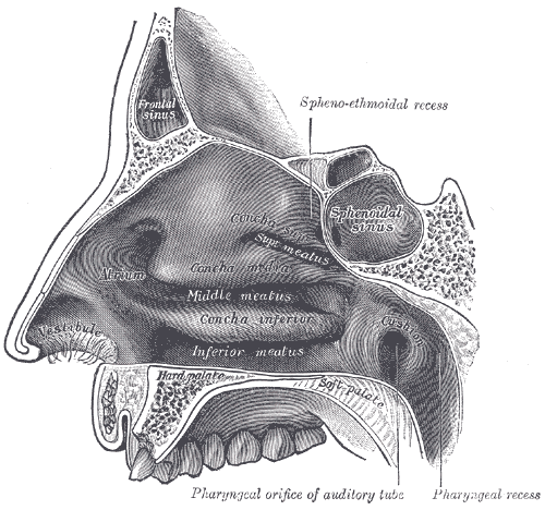
Posterior nasal apertures 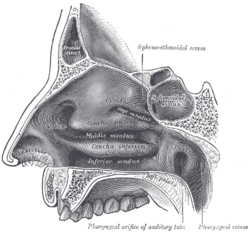
Lateral wall of nasal cavity. 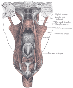
Dissection of the muscles of the palate from behind. (Choanae visible at center top.) Gray's subject #47 196 Choana (plural: Choanae; from Greek χοάνη "funnel") is the posterior nasal aperture.
The choanae are separated by the vomer.
Contents
Boundaries
It is the opening between the nasal cavity and the nasopharynx.
It is therefore not a structure but a space bounded as follows:
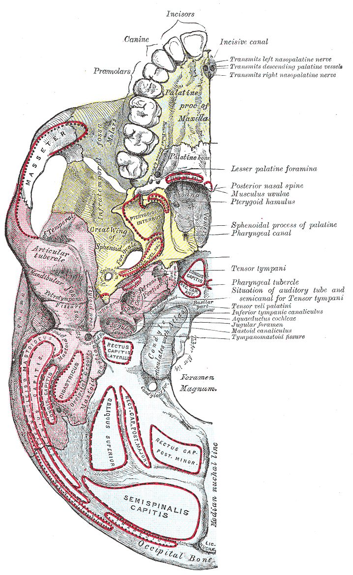
- anteriorly and inferiorly by the horizontal plate of palatine bone,
- superiorly and posteriorly by the sphenoid bone
- laterally by the medial pterygoid plates.
Etymology
The term is a latinization from the Greek "choanē" meaning funnel.
Choanae in different animals
The only animals with choana are the tetrapoda, and they could as well be called Choanata (they are also the only ones with a vomeronasal organ, which has an embryonic origin from the olfactory structure).
These internal nasal passages evolved while the vertebrates still lived in water. At this point they already needed to gulp air to get enough oxygen, and rather than open their jaws each time to do this, those mutants who acquired small openings to breathe through were more successful at living in the new environment.
Fish
Fish do not have choanae, instead they have two pairs of external nostrils: each with two tubes whose frontal openings lie close to the upper jaw, and the posterior openings further behind near the eyes. Whether choanae of tetrapods are homologous to the posterior nostrils or not has been debated. Reasons for dispute have been that the posterior nostril in its evolution into choanae would have to switch position relative to other anatomical features, i.e. a nerve.[1] Recent paleontologiclal findings support homology: a 400-million-year-old fossil lobe-finned fish called Kenichthys campbelli has something between a choana and the external nostrils seen on other fish, which makes it look like it has a cleft palate or cleft lip.[1] The reason seems to be that the posterior opening of the external nostrils has migrated into the mouth for some reason.
A similar evolution has taken place in lungfish. Here the inner nostrils have generally been accepted as homologous to the posterior nostrils, but the homology to true choanae as internal nostrils has been a matter of controversy. The fossil lungfish Diabolepis shows an intermediate stage between posterior and interior nostril and supports the independent origin of internal nostrils in the lungfish.[1]
Tetrapods
Similar migration is still seen in the tetrapod embryo, and can cause a baby to be born with a cleft palate. Why it should migrate is a mystery, since the nostrils would be useless as a breathing device before their final position inside the mouth. They could also already breathe air through their spiracles.
Tetrapods are also equipped with a lacrimal duct, or tear duct. How it evolved is not known, but it has an internal connection with the choana. It is possible that the choana started as a natural crack between maxilla and premaxilla because of an incomplete fusion in air-breathing animals. If this gap got wider and deeper with time, the frontal part of it would have to fuse together to avoid weakening the upper jaw, creating a small opening on the upper lip. Some more migrating, and this gap would meet the anterior pair of the external nasal openings. The posterior pair of the openings was then free to form the lacrimal duct if a migration caused them to come in contact with the eyes.
Choanae analogues in other animals and fossils
This would not have been the first time the jaws evolved some sort of opening. For instance, snakes have evolved a cleft in the lower jaw, allowing them to stick out their tongues without having to open the jaw. For an animal living in water, the formation of a paired cleft on the upper jaw would be quite logical. Terrestrial vertebrates would in any case need a way to breathe without needing to open their jaws each time.
Some fossil species are said to have both conventional external nostrils and a choana, but only more fossils will give a real answer to how the choanas evolved.
Lungfish and hagfishes
In addition to tetrapods, the lungfish has internal nostrils too. These seem to have a different origin than those of the tetrapods, and lungfish have no tear duct either.
Hagfishes have a single internal nostril that opens inside the mouth cavity, while Chimaerae have open canals that leads water from their external nostrils into their mouth and through their gills.
Additional images
-

Base of skull. Inferior surface.
References
Head and neck, upper RT: Nose (TA A06.1, TH H3.05.01, GA 10.992) External nose Ala of nose
nasal cartilages (of the septum, Greater alar, Lesser alar, Lateral nasal, Accessory nasal, Vomeronasal)Nasal cavity OpeningsLateral wallNasal concha/meati: Superior nasal concha · Middle nasal concha · Inferior nasal concha · Superior nasal meatus · Middle nasal meatus · Inferior nasal meatus
Sphenoethmoidal recess · Ethmoid bulla · Agger nasi · Ethmoidal infundibulum · Semilunar hiatus · Maxillary hiatusMedial wallParanasal sinuses Naso-pharynx Categories:

Wikimedia Foundation. 2010.