- Fuchs' dystrophy
-
Fuchs' dystrophy Classification and external resources 
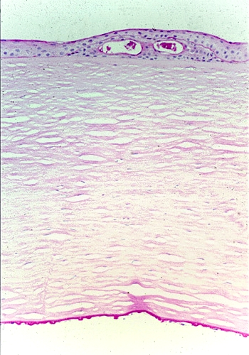
Fuchs corneal dystrophy. Light microscopic appearance of the cornea showing numerous excrescences (guttae) on the posterior surface of Descemet's membrane and the presence of cysts in the corneal epithelium beneath ectopically placed intraepithelial basement membrane. Periodic acid-Schiff stain. From a review by Klintworth, 2009.[1]ICD-10 H18.5 ICD-9 371.57 OMIM 136800 610158 DiseasesDB 31163 eMedicine article/1193591 MeSH D005642 Fuchs' dystrophy, also known as Fuchs' endothelial dystrophy, is a slowly progressing corneal disease that usually affects both eyes and is slightly more common in women than in men. Although doctors can often see early signs of Fuchs' dystrophy in people in their 30s and 40s, the disease rarely affects vision until people reach their 50s and 60s.
The condition was first described by Austrian Ernst Fuchs (1851–1930), after whom it is named.
Contents
Etiology
Fuchs’ endothelial dystrophy (FED) is a degenerative disorder of the corneal endothelium with accumulation of focal excrescences called guttae and thickening of Descemet’s membrane, leading to corneal edema and loss of vision. Corneal endothelial cells are the major “pump” cells of the cornea to allow for stromal clarity. In FED, Descemet’s membrane is grossly thickened with accumulation of abnormal wide-spaced collagen and numerous guttae. Corneal endothelial cells in end-stage FED are reduced in number and appear attenuated, causing progressive stromal edema. Progressive endothelial cell loss causes relative influx of aqueous humor into the cornea, leading to swelling (corneal stromal edema), which results in distorted vision. Eventually, the epithelium also becomes edematous, resulting in more severe visual impairment. Focal areas or blisters of epithelial edema ("bullae") may be particularly painful.
The inheritance of FED is autosomal dominant with genetic and environmental modifiers such as increased prevalence in the elderly and in females. Endothelial cell loss may be aggravated or accelerated by intraocular trauma or surgery. A common scenario involves excessive corneal swelling or edema following cataract surgery or other types of ocular surgery. Hence, patients with a history of Fuchs' dystrophy may be at a greater risk of corneal edema after ocular surgery as they have fewer functioning endothelial cells.
FED is classified into 4 stages, from early signs of guttae formation to end-stage subepithelial scarring. Diagnosis is made by biomicroscopic examination; other modalities, such as corneal pachymetry, confocal biomicroscopy, and specular microscopy can be used in conjunction.
Exact pathogenesis is unknown but factors include endothelial cell apoptosis, sex hormones, inflammation, and aqueous humor flow and composition. Mutations in collagen VIII, a major component of Descemet’s membrane secreted by endothelial cells, have been linked to the early-onset FED.[2]
Genes include:
Type OMIM Gene Locus FECD1 136800 COL8A2 1p34.3-p32.3 FECD4 610206 SLC4A11 20p13-p12 FECD6 189909 ZEB1 10p11.2 Signs and symptoms
At first, a person with Fuchs' dystrophy will awaken with blurred vision that will gradually clear during the day. This occurs because the cornea is normally thicker in the morning; it retains fluids during sleep that evaporate in the tear film while we are awake. As the disease worsens, this swelling will remain constant and reduce vision throughout the day.
Treatment
Medical management includes topical hypertonic saline, the use of a hairdryer to dehydrate the precorneal tear film, and therapeutic soft contact lenses. In using a hairdryer, the patient is instructed to hold a hairdryer at an arm's length or directed across the face, to dry out the epithelial blisters. This can be done two or three times a day. Definitive treatment, however, (especially with increased corneal edema) is surgical in the form of corneal transplantation, or penetrating keratoplasty (PKP).
Since 1998, new surgical modalities in the treatment of FED have been developed by Melles et al. in The Netherlands. These procedures, called posterior lamellar keratoplasty or endothelial keratoplasty, have been popularized as deep lamellar endothelial keratoplasty (DLEK) and Descemet’s stripping with endothelial keratoplasty (DSEK). DLEK and DSEK avoid the surgical complications of PKP such as wound dehiscence and infections and high postoperative astigmatism. Since 2004, DSEK has become the dominant procedure because it is technically much easier for the surgeon compared to DLEK or PKP. Improved surgical instrumentation for DSEK, such as a DSEK graft injector will become available shortly (2008). This could allow faster recovery for patients because of the ability to perform DSEK through very small (3 mm) sutureless incisions.
Recently, endothelial keratoplasty has been further refined to Descemet Membrane Endothelial Keratoplasty (DMEK), in which only a donor Descemet membrane and its endothelium is transplanted. With DMEK, 90% of cases achieve a best spectacle corrected visual acuity 20/40 or better, and 60% of cases 20/25 or better within 1–3 months.
More speculative future directions in the treatment of FED include in vitro expansion of human corneal endothelial cells for transplantation, artificial corneas and genetic modification.
See also
- Fuchs heterochromic iridocyclitis (a disease of the iris)
- Ocular straylight
References
- ^ Klintworth GK (2009). "Corneal dystrophies". Orphanet J Rare Dis 4: 7. doi:10.1186/1750-1172-4-7. PMC 2695576. PMID 19236704. http://www.ojrd.com/content/4//7.
- ^ Gottsch JD, Sundin OH, Liu SH, et al. (June 2005). "Inheritance of a novel COL8A2 mutation defines a distinct early-onset subtype of fuchs corneal dystrophy". Invest. Ophthalmol. Vis. Sci. 46 (6): 1934–9. doi:10.1167/iovs.04-0937. PMID 15914606. http://www.iovs.org/cgi/pmidlookup?view=long&pmid=15914606.
External links
- Facts About the Cornea and Corneal Disease The National Eye Institute (NEI)
- Descemet Membrane Endothelial Keratoplasty
- Summary of scientific literature
- The Corneal Dystrophy Foundation
Types of human corneal dystrophy (H18.5, 371.5) Epithelial and Subepithelial Epithelial basement membrane dystrophy (OMIM 121820), called a corneal dystrophy but in reality this condition is not inherited in the majority of cases, representing a non-specific reaction to a variety of corneal insults. · Subepithelial mucinous corneal dystrophy · Meesmann juvenile epithelial corneal dystrophy (MECD, Stocker-Holt dystrophy, OMIM 122100) · Lisch epithelial dystrophy · Gelatinous drop-like corneal dystrophyBowman layer Reis-Bucklers corneal dystrophy (CDB1) aka. Granular corneal dystrophy type III · Thiel-Behnke dystrophy (CDB2)Stroma Lattice corneal dystrophy type I · Lattice corneal dystrophy type II · Granular corneal dystrophy type I · Granular corneal dystrophy type II · Also Granular corneal dystrophy type III see Reis-Bucklers corneal dystrophy above · Macular corneal dystrophy · Schnyder corneal dystrophy · Congenital stromal dystrophy (CSCD) · Fleck dystrophy · Posterior amorphous corneal dystrophyDescemet membrane and Endothelial Fuchs' dystrophy · Posterior polymorphous dystrophy type 1 · Posterior polymorphous dystrophy type 2 · Posterior polymorphous dystrophy type 3 · Congenital endothelial dystrophy type 1 (CHED1) · Congenital endothelial dystrophy type 2 (CHED2) · X-linked endothelial corneal dystrophyM: EYE
anat(g/a/p)/phys/devp/prot
noco/cong/tumr, epon
proc, drug(S1A/1E/1F/1L)
Genetic disorder, extracellular: scleroprotein disease (excluding laminin and keratin) Collagen disease COL1: Osteogenesis imperfecta · Ehlers–Danlos syndrome, types 1, 2, 7
COL2: Hypochondrogenesis · Achondrogenesis type 2 · Stickler syndrome · Marshall syndrome · Spondyloepiphyseal dysplasia congenita · Spondyloepimetaphyseal dysplasia, Strudwick type · Kniest dysplasia (see also C2/11)
COL3: Ehlers–Danlos syndrome, types 3 & 4 (Sack–Barabas syndrome)
COL4: Alport syndrome
COL5: Ehlers–Danlos syndrome, types 1 & 2
COL6: Bethlem myopathy · Ullrich congenital muscular dystrophy
COL7: Epidermolysis bullosa dystrophica · Recessive dystrophic epidermolysis bullosa · Bart syndrome · Transient bullous dermolysis of the newborn
COL8: Fuchs' dystrophy 1
COL9: Multiple epiphyseal dysplasia 2, 3, 6
COL10: Schmid metaphyseal chondrodysplasia
COL11: Weissenbacher–Zweymüller syndrome · Otospondylomegaepiphyseal dysplasia (see also C2/11)
COL17: Bullous pemphigoidLaminin Junctional epidermolysis bullosa · Laryngoonychocutaneous syndromeOther Congenital stromal corneal dystrophy · Raine syndrome · Urbach–Wiethe disease · TECTA (DFNA8/12, DFNB21)Genetic disorder, membrane: Solute carrier disorders 1-10 SLC1A3 (Episodic ataxia 6) · SLC2A1 (De Vivo disease) · SLC2A5 (Fructose malabsorption) · SLC2A10 (Arterial tortuosity syndrome) · SLC3A1 (Cystinuria) · SLC4A1 (Hereditary spherocytosis 4/Hereditary elliptocytosis 4) · SLC4A11 (Congenital endothelial dystrophy type 2, Fuchs' dystrophy 4) · SLC5A1 (Glucose-galactose malabsorption) · SLC5A2 (Renal glycosuria) · SLC5A5 (Thyroid dyshormonogenesis type 1) · SLC6A19 (Hartnup disease) · SLC7A7 (Lysinuric protein intolerance) · SLC7A9 (Cystinuria)11-20 SLC11A1 (Crohn's disease) · SLC12A3 (Gitelman syndrome) · SLC16A1 (HHF7) · SLC16A2 (Allan–Herndon–Dudley syndrome) · SLC17A5 (Salla disease) · SLC17A8 (DFNA25)21-40 see also solute carrier family
B structural (perx, skel, cili, mito, nucl, sclr) · DNA/RNA/protein synthesis (drep, trfc, tscr, tltn) · membrane (icha, slcr, atpa, abct, othr) · transduction (iter, csrc, itra), trfkGenetic disorder, protein biosynthesis: Transcription factor/coregulator deficiencies (1) Basic domains 1.2: Feingold syndrome · Saethre-Chotzen syndrome
1.3: Tietz syndrome(2) Zinc finger
DNA-binding domains2.1 (Intracellular receptor): Thyroid hormone resistance · Androgen insensitivity syndrome (PAIS, MAIS, CAIS) · Kennedy's disease · PHA1AD pseudohypoaldosteronism · Estrogen insensitivity syndrome · X-linked adrenal hypoplasia congenita · MODY 1 · Familial partial lipodystrophy 3 · SF1 XY gonadal dysgenesis
2.2: Barakat syndrome · Tricho–rhino–phalangeal syndrome
2.3: Greig cephalopolysyndactyly syndrome/Pallister-Hall syndrome · Denys–Drash syndrome · Duane-radial ray syndrome · MODY 7 · MRX 89 · Townes–Brocks syndrome · Acrocallosal syndrome · Myotonic dystrophy 2
2.5: Autoimmune polyendocrine syndrome type 1(3) Helix-turn-helix domains 3.1: ARX (Ohtahara syndrome, Lissencephaly X2) · HLXB9 (Currarino syndrome) · HOXD13 (SPD1 Synpolydactyly) · IPF1 (MODY 4) · LMX1B (Nail–patella syndrome) · MSX1 (Tooth and nail syndrome, OFC5) · PITX2 (Axenfeld syndrome 1) · POU4F3 (DFNA15) · POU3F4 (DFNX2) · ZEB1 (Posterior polymorphous corneal dystrophy 3, Fuchs' dystrophy 3) · ZEB2 (Mowat-Wilson syndrome)
3.2: PAX2 (Papillorenal syndrome) · PAX3 (Waardenburg syndrome 1&3) · PAX4 (MODY 9) · PAX6 (Gillespie syndrome, Coloboma of optic nerve) · PAX8 (Congenital hypothyroidism 2) · PAX9 (STHAG3)
3.3: FOXC1 (Axenfeld syndrome 3, Iridogoniodysgenesis, dominant type) · FOXC2 (Lymphedema–distichiasis syndrome) · FOXE1 (Bamforth–Lazarus syndrome) · FOXE3 (Anterior segment mesenchymal dysgenesis) · FOXF1 (ACD/MPV) · FOXI1 (Enlarged vestibular aqueduct) · FOXL2 (Premature ovarian failure 3) · FOXP3 (IPEX)
3.5: IRF6 (Van der Woude syndrome, Popliteal pterygium syndrome)(4) β-Scaffold factors
with minor groove contacts4.2: Hyperimmunoglobulin E syndrome
4.3: Holt-Oram syndrome · Li-Fraumeni syndrome · Ulnar–mammary syndrome
4.7: Campomelic dysplasia · MODY 3 · MODY 5 · SF1 (SRY XY gonadal dysgenesis, Premature ovarian failure 7) · SOX10 (Waardenburg syndrome 4c, Yemenite deaf-blind hypopigmentation syndrome)
4.11: Cleidocranial dysostosis(0) Other transcription factors 0.6: Kabuki syndromeUngrouped Transcription coregulators Categories:- Disorders of sclera and cornea
- Collagen disease
Wikimedia Foundation. 2010.