- Retinal detachment
-
Retinal detachment Classification and external resources 
Slit lamp photograph showing retinal detachment.ICD-10 H33 ICD-9 361 eMedicine oph/504 MeSH D012163 Retinal detachment is a disorder of the eye in which the retina peels away from its underlying layer of support tissue. Initial detachment may be localized, but without rapid treatment the entire retina may detach, leading to vision loss and blindness. It is a medical emergency.[1]
The retina is a thin layer of light sensitive tissue on the back wall of the eye. The optical system of the eye focuses light on the retina much like light is focused on the film in a camera. The retina translates that focused image into neural impulses and sends them to the brain via the optic nerve. Occasionally, posterior vitreous detachment, injury or trauma to the eye or head may cause a small tear in the retina. The tear allows vitreous fluid to seep through it under the retina, and peel it away like a bubble in wallpaper.
Contents
Types
- Rhegmatogenous retinal detachment – A rhegmatogenous retinal detachment occurs due to a break in the retina that allows fluid to pass from the vitreous space into the subretinal space between the sensory retina and the retinal pigment epithelium. Retinal breaks are divided into three types - holes, tears and dialyses. Holes form due to retinal atrophy especially within an area of lattice degeneration. Tears are due to vitreoretinal traction. Dialyses which are very peripheral and circumferential may be either tractional or atrophic, the atrophic form most often occurring as idiopathic dialysis of the young.
- Exudative, serous, or secondary retinal detachment – An exudative retinal detachment occurs due to inflammation, injury or vascular abnormalities that results in fluid accumulating underneath the retina without the presence of a hole, tear, or break. In evaluation of retinal detachment it is critical to exclude exudative detachment as surgery will make the situation worse, not better. Although rare, exudative retinal detachment can be caused by the growth of a tumor on the layers of tissue beneath the retina, namely the choroid. This cancer is called a choroidal melanoma.
- Tractional retinal detachment – A tractional retinal detachment occurs when fibrous or fibrovascular tissue, caused by an injury, inflammation or neovascularization, pulls the sensory retina from the retinal pigment epithelium.
A minority of retinal detachments result from trauma, including blunt blows to the orbit, penetrating trauma, and concussions to the head. A retrospective Indian study of more than 500 cases of rhegmatogenous detachments found that 11% were due to trauma, and that gradual onset was the norm, with over 50% presenting more than one month after the inciting injury.[2]
Frequency
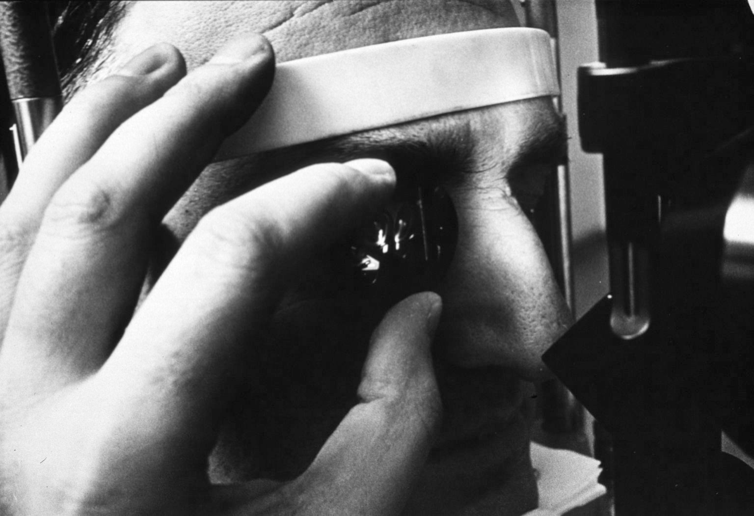
 A physician using a "three-mirror glass" to diagnose retinal detachment
The incidence of retinal detachment in otherwise normal eyes is around 5 new cases in 100,000 persons per year.[3] Detachment is more frequent in middle-aged or elderly populations, with rates of around 20 in 100,000 per year.[4] The lifetime risk in normal individuals is about 1 in 300.[5]
- Retinal detachment is more common in people with severe myopia (above 5–6 diopters), in whom the retina is more thinly stretched. In such patients, lifetime risk rises to 1 in 20.[6] About two-thirds of cases of retinal detachment occur in myopics. Myopic retinal detachment patients tend to be younger than non-myopic ones.
- Retinal detachment is more frequent after surgery for cataracts. The estimated long-term prevalence of retinal detachment after cataract surgery is in the range of 5 to 16 per 1000 cataract operations, [7] but is much higher in patients who are highly myopic, with a prevalence of up to 7% being reported in one study.[8] One study found that the probability of experiencing retinal detachment within 10 years of cataract surgery may be about 5 times higher than in the absence of treatment.[9]
- Tractional retinal detachments can also occur in patients with proliferative diabetic retinopathy [10] or those with proliferative retinopathy of sickle cell disease.[11] In proliferative retinopathy, abnormal blood vessels (neovascularization) grow within the retina and extend into the vitreous. In advanced disease, the vessels can pull the retina away from the back wall of the eye, leading to tractional retinal detachment.
Although retinal detachment usually occurs in just one eye, there is a 15% chance of it developing in the other eye, and this risk increases to 25–30% in patients who have had cataracts extracted from both eyes.[6]
Symptoms
A retinal detachment is commonly preceded by a posterior vitreous detachment which gives rise to these symptoms:
- flashes of light (photopsia) – very brief in the extreme peripheral (outside of center) part of vision
- a sudden dramatic increase in the number of floaters
- a ring of floaters or hairs just to the temporal side of the central vision
- a slight feeling of heaviness in the eye
Although most posterior vitreous detachments do not progress to retinal detachments, those that do produce the following symptoms:
- a dense shadow that starts in the peripheral vision and slowly progresses towards the central vision
- the impression that a veil or curtain was drawn over the field of vision
- straight lines (scale, edge of the wall, road, etc.) that suddenly appear curved (positive Amsler grid test)
- central visual loss
(None of this is to be confused with the broken retina which is generally the tearing of muscle and nerve behind the eye)
Treatment of Rhegmatogenous Retinal Detachment
There are several methods of treating a detached retina, each of which depends on finding and closing the breaks that have formed in the retina. All three of the procedures follow the same three general principles:
- Find all retinal breaks
- Seal all retinal breaks
- Relieve present (and future) vitreoretinal traction
- Cryopexy and Laser Photocoagulation
- Cryotherapy (freezing) or laser photocoagulation are occasionally used alone to wall off a small area of retinal detachment so that the detachment does not spread.
- Scleral buckle surgery
- Scleral buckle surgery is an established treatment in which the eye surgeon sews one or more silicone bands (bands, tyres) to the sclera (the white outer coat of the eyeball). The bands push the wall of the eye inward against the retinal hole, closing the break or reducing fluid flow through it and reducing the effect of vitreous traction thereby allowing the retina to re-attach. Cryotherapy (freezing) is applied around retinal breaks prior to placing the buckle. Often subretinal fluid is drained as part of the buckling procedure. The buckle remains in situ. The most common side effect of a scleral operation is myopic shift. That is, the operated eye will be more short sighted after the operation. Radial scleral buckle is indicated to U-shaped tears or Fishmouth tears and posterior breaks. Circumferential scleral buckle indicated to multiple breaks, anterior breaks and wide breaks. Encircling buckles indicated to breaks more than 2 quadrant of retinal area, lattice degeration located on more than 2 quadrant of retinal area, undetectable breaks, and proliferative vitreous retinopathy.
- Pneumatic retinopexy
- This operation is generally performed in the doctor's office under local anesthesia. It is another method of repairing a retinal detachment in which a gas bubble (SF6 or C3F8 gas) is injected into the eye after which laser or freezing treatment is applied to the retinal hole. The patient's head is then positioned so that the bubble rests against the retinal hole. Patients may have to keep their heads tilted for several days to keep the gas bubble in contact with the retinal hole. The surface tension of the air/water interface seals the hole in the retina, and allows the retinal pigment epithelium to pump the subretinal space dry and suck the retina back into place. This strict positioning requirement makes the treatment of the retinal holes and detachments that occurs in the lower part of the eyeball impractical. This procedure is usually combined with cryopexy or laser photocoagulation.
- Vitrectomy
- Vitrectomy is an increasingly used treatment for retinal detachment. It involves the removal of the vitreous gel and is usually combined with filling the eye with either a gas bubble (SF6 or C3F8 gas) or silicon oil. An advantage of using gas in this operation is that there is no myopic shift after the operation and gas is absorbed within a few weeks. Silicon oil (PDMS), if filled needs to be removed after a period of 2–8 months depending on surgeon's preference. Silicon oil is more commonly used in cases associated with proliferative vitreo-retinopathy (PVR). A disadvantage is that a vitrectomy always leads to more rapid progression of a cataract in the operated eye. In many places vitrectomy is the most commonly performed operation for the treatment of retinal detachment.
Results of Surgery
85 percent of cases will be successfully treated with one operation with the remaining 15 percent requiring 2 or more operations. After treatment patients gradually regain their vision over a period of a few weeks, although the visual acuity may not be as good as it was prior to the detachment, particularly if the macula was involved in the area of the detachment. However, if left untreated, total blindness could occur in a matter of days.
Risk factors and prevention
History of cataract surgery is an important risk factor for rhegmatogenous retinal detachment, which can manifest long after the operation has been completed. The risk is increased when there are complications during cataract surgery.
Retinal detachment can be mitigated in some cases when the warning signs[12] are caught early. The most effective means of prevention and risk reduction is through education of the initial signs, and encouragement for people to seek ophthalmic medical attention if they suffer from symptoms suggestive of a posterior vitreous detachment.[13] Early examination allows detection of retinal tears which can be treated with laser or cryotherapy. This reduces the risk of retinal detachment in those who have tears from around 1:3 to 1:20. For this reason, the governing bodies in some sports require regular eye examination.
Trauma-related cases of retinal detachment can occur in high-impact sports (eg boxing, karate, kickboxing, American football) or in high speed sports (eg automobile racing, sledding). Although some doctors recommend avoiding activities that increase pressure in the eye, including diving and skydiving, there is little evidence to support this recommendation, especially in the general population. Nevertheless, ophthalmologists generally advise patients with high degrees of myopia to try to avoid exposure to activities that have the potential for trauma, increase pressure on or within the eye itself, or include rapid acceleration and deceleration.
Intraocular pressure spikes occur during any activity accompanied by the Valsalva maneuver, including weightlifting.[14] An epidemiological study suggests that heavy manual lifting at work may be associated with increased risk of rhegmatogenous retinal detachment.[15][16] In this study, obesity also appeared to increase the risk of retinal detachment.
See also
References
- ^ "Retinal detachment". MedlinePlus Medical Encyclopedia. National Institutes of Health. 2005. http://www.nlm.nih.gov/medlineplus/ency/article/001027.htm. Retrieved 2006-07-18.
- ^ Shukla Manoj, Ahuja OP, Jamal Nasir. "Epidemiological study of nontraumatic phakic rhegmatogenous retinal detachment". Indian J Ophthalmol 1986;34:29–32.
- ^ Ivanisević M, Bojić L, Eterović D (2000). "Epidemiological study of nontraumatic phakic rhegmatogenous retinal detachment". Ophthalmic Res. 32 (5): 237–9. doi:10.1159/000055619. PMID 10971186.
- ^ Li X; Beijing Rhegmatogenous Retinal Detachment Study Group (2003). "Incidence and epidemiological characteristics of rhegmatogenous retinal detachment in Beijing, China". Ophthalmology 110 (12): 2413–7. doi:10.1016/S0161-6420(03)00867-4. PMID 14644727.
- ^ "Evaluation and Management of Suspected Retinal Detachment - April 1, 2004 - American Family Physician". http://www.aafp.org/afp/20040401/1691.html. Retrieved 2007-06-04.
- ^ a b "eMedicine – Retinal Detachment : Article by Gregory Luke Larkin, MD, MSPH, MSEng, FACEP". http://www.emedicine.com/emerg/topic504.htm. Retrieved 2007-06-04.
- ^ Ramos M, Kruger EF, Lashkari K (2002). "Biostatistical analysis of pseudophakic and aphakic retinal detachments". Seminars in ophthalmology 17 (3–4): 206–13. doi:10.1076/soph.17.3.206.14784. PMID 12759852.
- ^ Hyams SW, Bialik M, Neumann E (1975). "Myopia-aphakia. I. Prevalence of retinal detachment". The British journal of ophthalmology 59 (9): 480–2. doi:10.1136/bjo.59.9.480. PMC 1042658. PMID 1203233. http://www.pubmedcentral.nih.gov/articlerender.fcgi?tool=pmcentrez&artid=1042658.
- ^ J.A. Rowe, J.C. Erie, K.H. Baratz et al. (1999). "Retinal detachment in Olmsted County, Minnesota, 1976 through 1995". Ophthalmology 106 (1): 154–159. doi:10.1016/S0161-6420(99)90018-0. PMID 9917797.
- ^ "Diabetic Retinopathy: Retinal Disorders: Merck Manual Home Health Handbook". http://www.merckmanuals.com/home/eye_disorders/retinal_disorders/diabetic_retinopathy.html. Retrieved 2007-06-04.
- ^ "IU Opt Online CE: Retinal Vascular Disease: Sickle Cell Retinopathy". http://www.opt.indiana.edu/ce/retvasdz/sickle.htm. Retrieved 2007-06-04.
- ^ Retinal Detachment - NHS Choices
- ^ Byer NE (1994). "Natural history of posterior vitreous detachment with early management as the premier line of defense against retinal detachment". Ophthalmology 101 (9): 1503–13; discussion 1513–4. PMID 8090453.
- ^ Dickerman RD, Smith GH, Langham-Roof L, McConathy WJ, East JW, Smith AB (1999). "Intra-ocular pressure changes during maximal isometric contraction: does this reflect intra-cranial pressure or retinal venous pressure?". Neurol. Res. 21 (3): 243–6. PMID 10319330.
- ^ Mattioli S, De Fazio R, Buiatti E, Truffelli D, Zanardi F, Curti S, Cooke RM, Baldasseroni A, Miglietta B, Bonfiglioli R, Tassinari G, Violante FS (2008). "Physical exertion (lifting) and retinal detachment among people with myopia". Epidemiology 19 (6): 868–71. doi:10.1097/EDE.0b013e318187a7da. PMID 18854710.
- ^ Mattioli, S.; Curti, S.; De Fazio, R.; Farioli, A.; Cooke, R. M. T.; Zanardi, F.; Violante, F. S. (2009). "Risk Factors for Retinal Detachment". Epidemiology 20 (3): 465–466. doi:10.1097/EDE.0b013e31819f1b17. PMID 19363359.
External links
- The Vitreoretinal Company Manufacturer of Medical Devices for Retinal Detachment Surgery (MIRA).
- Retinal Detachment Resource Guide from the National Eye Institute (NEI).
- Overview of retinal detachment from eMedicine
- Guidelines from the American Academy of Family Physicians
- Retinal Clinic Rushabh Eye
- Retinal detachment information from WebMD
- Retinal detachment information from the Merck Manual
- A Diary of Retinal Detachment
- Royal National Institute for the Blind(UK )Site
- Detached Retina and The Treatment Required
- Engineering a new tool for observing the retina and preventing detachment Ingenia, March 2007
- Steven Fisher's Retinal Cell Biology lab at the University of California, Santa Barbara
- Advice from someone who has experienced Retinal Detachment
- 3D animation video explanation from youtube
Categories:- Medical emergencies
- Disorders of choroid and retina

Wikimedia Foundation. 2010.