- Nodule of vermis
-
Brain: Nodule of vermis 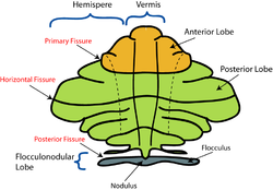
Schematic representation of the major anatomical subdivisions of the cerebellum. Superior view of an "unrolled" cerebellum, placing the vermis in one plane. 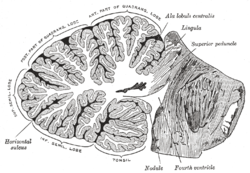
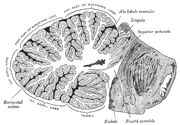
Sagittal section of the cerebellum, near the junction of the vermis with the hemisphere. (Nodule labeled at bottom right.) Latin nodulus vermis Gray's subject #187 791 NeuroNames hier-678 The nodule (nodular lobe), or anterior end of the inferior vermis, abuts against the roof of the fourth ventricle, and can only be distinctly seen after the cerebellum has been separated from the medulla oblongata and pons.
On either side of the nodule is a thin layer of white substance, named the posterior medullary velum.
It is semilunar in form, its convex border being continuous with the white substance of the cerebellum; it extends on either side as far as the flocculus.
External links
Human brain, rhombencephalon, metencephalon: cerebellum (TA 14.1.07, GA 9.788) Surface anatomy LobesMedial/lateralVermis: anterior (Central lobule, Culmen, Lingula) · posterior (Folium, Tuber, Uvula) · Vallecula of cerebellum
Hemisphere: anterior (Alar central lobule) · posterior (Biventer lobule, Cerebellar tonsil)Grey matter Molecular layer (Stellate cell, Basket cell)
Purkinje cell layer (Purkinje cell, Bergmann glia cell = Golgi epithelial cell)
Granule cell layer (Golgi cell, Granule cell, Unipolar brush cell)
Fibers: Mossy fibers · Climbing fiber · Parallel fiberWhite matter InternalPedunclesInferior (medulla): Dorsal spinocerebellar tract · Olivocerebellar tract · Cuneocerebellar tract · Juxtarestiform body (Vestibulocerebellar tract)
Middle (pons): Pontocerebellar fibers
Superior (midbrain): Ventral spinocerebellar tract · Dentatothalamic tract · Trigeminocerebellar fibersCategories:- Brain
- Neuroscience stubs

Wikimedia Foundation. 2010.