- Reticular formation
-
Brain: reticular formation 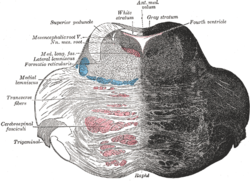
Coronal section of the pons, at its upper part. (Formatio reticularis labeled at left.) 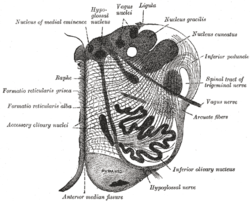
Section of the medulla oblongata at about the middle of the olive. (Formatio reticularis grisea and formatio reticularis alba labeled at left.) Latin formatio reticularis Gray's subject #187 784 NeuroNames ancil-225 MeSH Reticular+Formation The reticular formation is a part of the brain that is involved in actions such as awaking/sleeping cycle, and filtering incoming stimuli to discriminate irrelevant background stimuli.[1] It is essential for governing some of the basic functions of higher organisms, and is one of the phylogenetically oldest portions of the brain.
Contents
Functions
The reticular formation consists of more than 100 small neural networks, with varied functions including the following:
1. Somatic motor control - Some motor neurons send their axons to the reticular formation nuclei, giving rise to the reticulospinal tracts of the spinal cord. These tracts function in maintaining tone, balance, and posture--especially during body movements. The reticular formation also relays eye and ear signals to the cerebellum so that the cerebellum can integrate visual, auditory, and vestibular stimuli in motor coordination. Other motor nuclei include gaze centers, which enable the eyes to track and fixate objects, and central pattern generators, which produce rhythmic signals to the muscles of breathing and swallowing.
2. Cardiovascular control - The reticular formation includes the cardiac and vasomotor centers of the medulla oblongata.
3. Pain modulation - The reticular formation is one means by which pain signals from the lower body reach the cerebral cortex. It is also the origin of the descending analgesic pathways. The nerve fibers in these pathways act in the spinal cord to block the transmission of some pain signals to the brain.
4. Sleep and consciousness - The reticular formation has projections to the thalamus and cerebral cortex that allow it to exert some control over which sensory signals reach the cerebrum and come to our conscious attention. It plays a central role in states of consciousness like alertness and sleep. Injury to the reticular formation can result in irreversible coma.
5. Habituation - This is a process in which the brain learns to ignore repetitive, meaningless stimuli while remaining sensitive to others. A good example of this is when a person can sleep through loud traffic in a large city, but is awakened promptly due to the sound of an alarm or crying baby. Reticular formation nuclei that modulate activity of the cerebral cortex are called the reticular activating system or extrathalamic control modulatory system.
Pathology
Mass lesions in the brain stem cause severe alterations in level of consciousness such as coma due to their effects on the reticular formation.[4] Bilateral damage to the reticular formation of the midbrain may lead to a coma or death.[5]
Lesions in the reticular formation have been found in the brains of people who have post-polio syndrome, and some imaging studies have shown abnormal activity in the area in people with chronic fatigue syndrome, indicating a high likelihood that damage to the reticular formation is responsible for the fatigue experienced with these syndromes.
History and etymology
The term "reticular formation" was coined in the late 19th century, coinciding with Ramon y Cajal’s neuron doctrine. Allan Hobson states in his book The Reticular Formation revisited that he thought the name is an etymological vestige from the fallen era of the aggregate field theory in the neural sciences. The term reticulum means a netlike structure, which is what the Reticular Formation appears to be at first glance. It has been described as being either too complex to study or an undifferentiated part of the brain with no organization at all. Eric Kandel even describes the reticular formation as being organized in a similar manner to the intermediate gray matter of the spinal cord. This chaotic, loose, and intricate form of organization is what has turned off many researchers from looking farther into this mysterious area of the brain that seems to be at the crux of basic neurological and behavioral functions of the human being[citation needed]. The cells lack clear ganglionic boundaries, but do have clear functional organizations and distinct cell types.
The term reticular formation is seldom used any longer except to speak in generalities. Modern anatomy, or neuroscience articles, usually refer to the individual nuclei that comprise the reticular formation.
Structure
 A cross section of the lower part of the pons showing the pontine reticular formation labeled as #8
The reticular formation has been functionally cleaved both sagittally and coronally.
- The original functional differentiation was a division of caudal and rostral, this was based upon the observation that the lesioning of the rostral reticular formation induces a hypersomnia in the cat brain. In contrast, lesioning of the more caudal portion of the reticular formation produces insomnia in cats. This study has led to the idea that the caudal portion inhibits the rostral portion of the reticular formation.
- Sagittal division reveals more morphological distinctions. The raphe nuclei form a ridge in the middle of the reticular formation, and, directly to its periphery, there is a division called the medial reticular formation. The medial RF is large and has long ascending and descending fibers, and is surrounded by the lateral reticular formation. The lateral RF is close to the motor nuclei of the cranial nerves, and mostly mediates their function.
Medial and lateral reticular formation
The medial reticular formation and lateral reticular formation are two columns of neuronal nuclei with ill-defined boundaries, which go up through the medulla and into the mesencephalon (midbrain). The nuclei can only be teased out by function, cell type, and projections of efferent or afferent nature.
References
- ^ http://thebrain.mcgill.ca/flash/a/a_11/a_11_cr/a_11_cr_cyc/a_11_cr_cyc.html
- ^ http://biology.about.com/library/organs/brain/blreticular.htm
- ^ Saladin, Kenneth S. Anatomy & physiology the unity of form and function. Dubuque: McGraw-Hill, 2009. Print.
- ^ Tindall SC (1990). "Level of consciousness". In Walker HK, Hall WD, Hurst JW. Clinical Methods: The History, Physical, and Laboratory Examinations and the Ana banana. Butterworth Publishers. http://www.ncbi.nlm.nih.gov/bookshelf/br.fcgi?book=cm&partid=380. Retrieved 2008-07-04.
- ^ The Human Brain: An Introduction to its Functional Anatomy 5th ed by J Nolte chpt 11 pp. 262–290
See also
Additional images
-
Deep dissection of brain-stem. Ventral view.
-

The formatio reticularis of the medulla oblongata, shown by a transverse section passing through the middle of the olive.
External links
Anatomy of torso (primarily): the spinal cord (TA 14.1.02, GA 9.749) External, dorsal Posterior median sulcus · Posterolateral sulcusGrey matter/
Rexed laminaeI–VI: Posterior hornI: Marginal nucleus · II: Substantia gelatinosa of Rolando · III+IV: Nucleus proprius · Spinal lamina V · Spinal lamina VIVII: Lateral hornVIII–IX: Anterior hornX: OtherWhite matter somatic/
ascending
(blue)Posterior/PCML: touch: Gracile · Cuneate
Lateral: proprioception: Spinocerebellar (Dorsal, Ventral) · pain/temp: Spinothalamic (Lateral, Anterior) · Posterolateral (Lissauer) · Spinotectal
Spinoreticular tract · Spino-olivary tractmotor/
descending
(red)Lateral: Corticospinal (Lateral) · Ep (Rubrospinal, Olivospinal)
Anterior: Corticospinal (Anterior) · Ep (Vestibulospinal, Reticulospinal, Tectospinal)bothExternal, ventral Anterior median fissure · Anterolateral sulcusExternal, general Human brain: rhombencephalon, myelencephalon: medulla (TA A14.1.04, GA 9.767) Dorsal SurfacePosterior median sulcus · Posterolateral sulcus · Area postrema · Vagal trigone · Hypoglossal trigone · Medial eminenceafferent: GVA: VII,IX,X: Solitary/tract/Dorsal respiratory group · SVA: Gustatory nucleus · GSA: VIII-v (Lateral, Medial, Inferior)
efferent: GSE: XII · GVE: IX,X,XI: Ambiguus · SVE: X: Dorsal · IX: Inferior salivatory nucleusGrey: otherWhite: Sensory/ascendingWhite: Motor/descendingVentral White: Motor/descendingVentral respiratory group · Arcuate nucleus of medulla · Inferior olivary nucleus · Rostral ventromedial medullaSurfaceGrey: Raphe/
reticularReticular formation (Gigantocellular, Parvocellular, Ventral, Lateral, Paramedian) · Raphe nuclei (Obscurus, Magnus, Pallidus)Human brain, rhombencephalon, metencephalon: pons (TA A14.1.05.101–604, GA 9.785) Dorsal/
(tegmentum)SurfaceWhite: Sensory/ascendingTrapezoid body/VIII · Trigeminal lemniscus (Dorsal trigeminal tract, Ventral trigeminal tract) · Medial lemniscus · Lateral lemniscus
MLF, III, IV and VI: Vestibulo-oculomotor fibers
Anterior trigeminothalamic tract · Central tegmental tractWhite: Motor/descendingICP (Vestibulocerebellar tract)
MLF, III, IV and VI: Vestibulospinal tract (Medial vestibulospinal tract, Lateral vestibulospinal tract)Other greyVentral/
(base)White: Motor/descendingSurfaceBasilar sulcusOther grey: Raphe/
reticularCategories:- Brainstem


Wikimedia Foundation. 2010.