- Vagal trigone
-
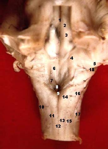
Brain: Vagal trigone 
Dissection of brain-stem. Dorsal view. 
Human caudal brainstem posterior view (Trigonum nervi vagi is #7) Latin trigonum nervi vagi Gray's subject #187 781 NeuroNames hier-631 The cells of the dorsal nucleus are spindle-shaped, like those of the posterior column of the spinal cord, and the nucleus is usually considered as representing the base of the posterior column. It measures about 2 cm. in length, and in the lower, closed part of the medulla oblongata is situated behind the hypoglossal nucleus; whereas in the upper, open part it lies lateral to that nucleus, and corresponds to an eminence, named the vagal trigone (ala cinerea, not to be confused with tuberculum cinereum nor tuber cinereum), in the rhomboid fossa.
Additional images
-

Rhomboid fossa.
External links
This article was originally based on an entry from a public domain edition of Gray's Anatomy. As such, some of the information contained within it may be outdated.
Human brain: rhombencephalon, myelencephalon: medulla (TA A14.1.04, GA 9.767) Dorsal SurfacePosterior median sulcus · Posterolateral sulcus · Area postrema · Vagal trigone · Hypoglossal trigone · Medial eminenceafferent: GVA: VII,IX,X: Solitary/tract/Dorsal respiratory group · SVA: Gustatory nucleus · GSA: VIII-v (Lateral, Medial, Inferior)
efferent: GSE: XII · GVE: IX,X,XI: Ambiguus · SVE: X: Dorsal · IX: Inferior salivatory nucleusGrey: otherWhite: Sensory/ascendingWhite: Motor/descendingVentral White: Motor/descendingVentral respiratory group · Arcuate nucleus of medulla · Inferior olivary nucleus · Rostral ventromedial medullaSurfaceGrey: Raphe/
reticularVentricular system, rhombencephalon, met- and myel-: fourth ventricle (TA A14.1.05.701–726, GA 9.797) Roof (dorsal) rostral: Superior medullary velum (Frenulum)
caudal: Inferior medullary velum · Taenia of fourth ventricleFloor/rhomboid fossa (ventral) rostral (pons): Facial colliculus · Locus coeruleus
caudal (medulla}: Vagal trigone · Hypoglossal trigone · Area postrema · Obex
Medial eminence · Sulcus limitansApertures Other Tela chorioidea of fourth ventricle · FastigiumCategories:- Brainstem
- Neuroscience stubs
-


Wikimedia Foundation. 2010.