- Pectineus muscle
-
Pectineus 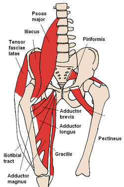
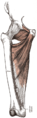
The pectineus and nearby muscles 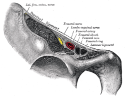
Structures passing behind the inguinal ligament. (Pectineus visible at bottom right.) Latin musculus pectineus Gray's subject #128 472 Origin pubis - superior pubic ramus Insertion Pectineal line of the Femur Artery Obturator artery Nerve Femoral nerve, sometimes obturator nerve Actions Thigh - flexion, adduction, medial rotation The pectineus muscle (from the Latin word pecten, meaning comb[1]) is a flat, quadrangular muscle, situated at the anterior part of the upper and medial aspect of the thigh.
It can be classified in the medial compartment of thigh[2] (when the function is emphasized) or the anterior compartment of thigh (when the nerve is emphasized).[3]
Contents
Origin and insertion
It arises from the pectineal line of the pubis and to a slight extent from the surface of bone in front of it, between the iliopectineal eminence and tubercle of the pubis, and from the fascia covering the anterior surface of the muscle; the fibers pass downward, backward, and lateralward, to be inserted into the pectineal line of the femur which leads from the lesser trochanter to the linea aspera.
Relations
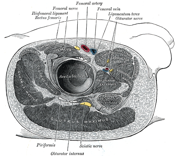
The pectineus is in relation by its anterior surface with the pubic portion of the fascia lata, which separates it from the femoral artery and vein and internal saphenous vein, and lower down with the profunda artery.
By its posterior surface with the capsule of the hip-joint, and with the obturator externus and adductor brevis, the obturator artery and vein being interposed.
By its external border with the psoas, the femoral artery resting upon the line of interval.
By its internal border with the outer edge of the adductor longus.
Obturator hernia is situated directly behind this muscle, which forms one of its coverings.
Action
It is one of the muscles primarily responsible for hip flexion. It also adducts and medially rotates the thigh.
The pectineus muscle is the most anterior adductor of the hip.
Innervation
Innervation is by the femoral nerve (L2 and L3) and occasionally (20% of the population) a branch of the obturator nerve called the accessory obturator nerve.
Additional images
-

Right hip bone. External surface.
-
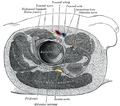
Structures surrounding right hip-joint.
-
Muscles of the iliac and anterior femoral regions.
-
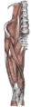
Deep muscles of the medial femoral region.
-
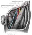
The left femoral triangle.
-
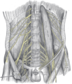
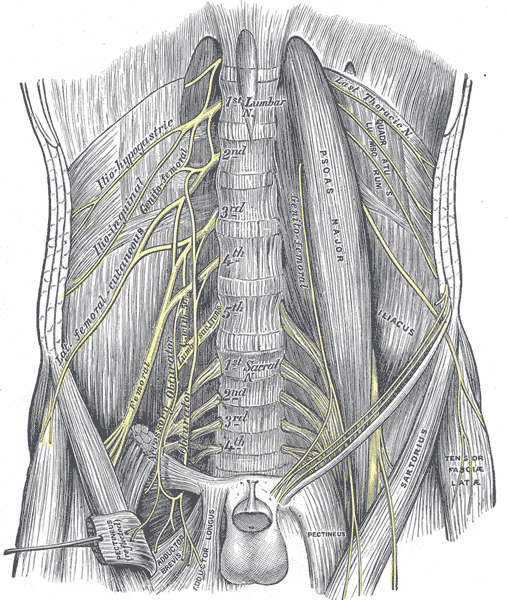
The lumbar plexus and its branches.
References
- ^ Mosby’s Medical, Nursing and Allied Health Dictionary, Fourth Edition, Mosby-Year Book Inc., 1994, p. 1177
- ^ Ellis, Harold; Susan Standring; Gray, Henry David (2005). Gray's anatomy: the anatomical basis of clinical practice. St. Louis, Mo: Elsevier Churchill Livingstone. p. 518. ISBN 0-443-07168-3.
- ^ medialthigh at The Anatomy Lesson by Wesley Norman (Georgetown University)
- ^ Wilson, Erasmus (1851). The anatomist's vade mecum: a system of human anatomy. p. 260. http://books.google.com/books?id=t2M1AV1aYWQC&pg=PA260.
This article was originally based on an entry from a public domain edition of Gray's Anatomy. As such, some of the information contained within it may be outdated.
External links
- -1301610416 at GPnotebook
- LUC pect
- SUNY Figs 12:02-05 - "Muscles of the anterior (extensor) compartment of the thigh."
- SUNY Figs 12:03-04 - "Deep muscles of the anterior thigh."
- Cross section at UV pelvis/pelvis-e12-15
List of muscles of lower limbs (TA A04.7, GA 4.465) ILIAC Region
/ ILIOPSOASBUTTOCKS THIGH /
compartmentsLEG/
Crus/
compartmentssuperficial · triceps surae (gastrocnemius, soleus, accessory soleus, Achilles tendon) · plantaris
deep · tarsal tunnel (flexor hallucis longus, flexor digitorum longus, tibialis posterior) · popliteusfibularis muscles (longus, brevis)FOOT DorsalPlantar1st layer (abductor hallucis, flexor digitorum brevis, abductor digiti minimi) · 2nd layer (quadratus plantae, lumbrical muscle) · 3rd layer (flexor hallucis brevis, adductor hallucis, flexor digiti minimi brevis) · 4th layer (dorsal interossei, plantar interossei)Categories:- Hip adductors
- Hip medial rotators
- Hip flexors
- Thigh muscles
- Muscle stubs
-


Wikimedia Foundation. 2010.