- Neuroglia
-
Neuroglia MeSH Neuroglia Code TA A14.0.00.005
TH H2.00.06.2.00001 Neuroglia of the brain shown by Golgi's method.
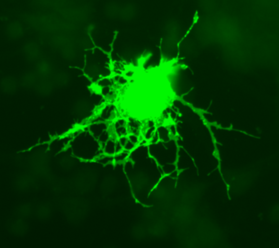
 Astrocytes can be identified in culture because, unlike other mature glia, they express glial fibrillary acidic protein.
 Glial cells in a rat brain stained with an antibody against GFAP.
Glial cells, sometimes called neuroglia or simply glia (Greek γλία, γλοία "glue"; pronounced in English either /gliːə/ or /glaɪə/), are non-neuronal cells that maintain homeostasis, form myelin, and provide support and protection for the brain's neurons. In the human brain, there is roughly one glia for every neuron with a ratio of about two neurons for every glia in the cerebral gray matter.[1]
As the Greek name implies, glia are commonly known as the glue of the nervous system; however, this is not fully accurate. Neuroscience currently identifies four main functions of glial cells: to surround neurons and hold them in place, to supply nutrients and oxygen to neurons, to insulate one neuron from another, and to destroy pathogens and remove dead neurons. For over a century, it was believed that they did not play any role in neurotransmission. That idea is now discredited;[2] they do modulate neurotransmission, although the mechanisms are not yet well understood.[2][3][4]
Contents
Functions
Some glial cells function primarily as the physical support for neurons. Others regulate the internal environment of the brain, especially the fluid surrounding neurons and their synapses, and nutrify neurons. During early embryogenesis glial cells direct the migration of neurons and produce molecules that modify the growth of axons and dendrites. Recent research[which?] indicates that glial cells of the hippocampus and cerebellum participate in synaptic transmission, regulate the clearance of neurotransmitters from the synaptic cleft, and release gliotransmitters such as ATP, which modulate synaptic function.
Glial cells are known to be capable of mitosis. By contrast, scientific understanding of whether neurons are permanently post-mitotic,[5] or capable of mitosis,[6] is still developing. In the past, glia had been considered[by whom?] to lack certain features of neurons. For example, glial cells were not believed to have chemical synapses or to release transmitters. They were considered to be the passive bystanders of neural transmission. However, recent studies have shown this to be untrue.[7]
For example, astrocytes are crucial in clearance of neurotransmitters from within the synaptic cleft, which provides distinction between arrival of action potentials and prevents toxic build-up of certain neurotransmitters such as glutamate (excitotoxicity). It is also thought that glia play a role in many neurological diseases, including Alzheimer's disease. Furthermore, at least in vitro, astrocytes can release gliotransmitter glutamate in response to certain stimulation. Another unique type of glial cell, the oligodendrocyte precursor cells or OPCs, have very well-defined and functional synapses from at least two major groups of neurons.[citation needed] The only notable differences between neurons and glial cells are neurons' possession of axons and dendrites, and capacity to generate action potentials.
Glia ought not to be regarded as "glue" in the nervous system as the name implies; rather, they are more of a partner to neurons.[8] They are also crucial in the development of the nervous system and in processes such as synaptic plasticity and synaptogenesis. Glia have a role in the regulation of repair of neurons after injury. In the CNS (Central Nervous System), glia suppress repair. Glial cells known as astrocytes enlarge and proliferate to form a scar and produce inhibitory molecules that inhibit regrowth of a damaged or severed axon. In the PNS (Peripheral Nervous System), glial cells known as Schwann cells promote repair. After axonal injury, Schwann cells regress to an earlier developmental state to encourage regrowth of the axon. This difference between PNS and CNS raises hopes for the regeneration of nervous tissue in the CNS. For example a spinal cord may be able to be repaired following injury or severance.
Types
Microglia
For more details on this topic, see Microglia.Microglia are like specialized macrophages capable of phagocytosis that protect neurons of the central nervous system.[9] They are derived from hematopoietic precursors rather than ectodermal tissue; they are commonly categorized as such because of their supportive role to neurons.
These cells comprise approximately 15% of the total cells of the central nervous system.[citation needed] They are found in all regions of the brain and spinal cord. Microglial cells are small relative to macroglial cells, with changing shapes and oblong nuclei. They are mobile within the brain and multiply when the brain is damaged. In the healthy central nervous system, microglia processes constantly sample all aspects of their environment (neurons, macroglia and blood vessels).
Macroglia
Location Name Description CNS Astrocytes The most abundant type of macroglial cell, astrocytes (also called astroglia) have numerous projections that anchor neurons to their blood supply. They regulate the external chemical environment of neurons by removing excess ions, notably potassium, and recycling neurotransmitters released during synaptic transmission. The current theory suggests that astrocytes may be the predominant "building blocks" of the blood-brain barrier. Astrocytes may regulate vasoconstriction and vasodilation by producing substances such as arachidonic acid, whose metabolites are vasoactive.
Astrocytes signal each other using calcium. The gap junctions (also known as electrical synapses) between astrocytes allow the messenger molecule IP3 to diffuse from one astrocyte to another. IP3 activates calcium channels on cellular organelles, releasing calcium into the cytoplasm. This calcium may stimulate the production of more IP3. The net effect is a calcium wave that propagates from cell to cell. Extracellular release of ATP, and consequent activation of purinergic receptors on other astrocytes, may also mediate calcium waves in some cases.
In general, there are two types of astrocytes, protoplasmic and fibrous, similar in function but distinct in morphology and distribution. Protoplasmic astrocytes have short, thick, highly branched processes and are typically found in gray matter. Fibrous astrocytes have long, thin, less branched processes and are more commonly found in white matter.
It has recently been shown that astrocyte activity is linked to blood flow in the brain, and that this is what is actually being measured in fMRI.[10]
CNS Oligodendrocytes Oligodendrocytes are cells that coat axons in the central nervous system (CNS) with their cell membrane forming a specialized membrane differentiation called myelin, producing the so-called myelin sheath. The myelin sheath provides insulation to the axon that allows electrical signals to propagate more efficiently.[11]
CNS Ependymal cells Ependymal cells, also named ependymocytes, line the cavities of the CNS and make up the walls of the ventricles. These cells create and secrete cerebrospinal fluid(CSF) and beat their cilia to help circulate the CSF and make up the Blood-CSF barrier. They are also thought to act as neural stem cells.[12]
CNS Radial glia Radial glia cells arise from neuroepithelial cells after the onset of neurogenesis. Their differentiation abilities are more restricted than those of neuroepithelial cells. In the developing nervous system, radial glia function both as neuronal progenitors and as a scaffold upon which newborn neurons migrate. In the mature brain, the cerebellum and retina retain characteristic radial glial cells. In the cerebellum, these are Bergmann glia, which regulate synaptic plasticity. In the retina, the radial Müller cell is the principal glial cell, and participates in a bidirectional communication with neurons.[13]
PNS Schwann cells Similar in function to oligodendrocytes, Schwann cells provide myelination to axons in the peripheral nervous system (PNS). They also have phagocytotic activity and clear cellular debris that allows for regrowth of PNS neurons.[14]
PNS Satellite cells Satellite glial cells are small cells that surround neurons in sensory, sympathetic and parasympathetic ganglia.[15] These cells help regulate the external chemical environment. Like astrocytes, they are interconnected by gap junctions and respond to ATP by elevating intracellular concentration of calcium ions. They are highly sensitive to injury and inflammation, and appear to contribute to pathological states, such as chronic pain.[16]
PNS Enteric glial cells Are found in the intrinsic ganglia of the digestive system.They are thought to have many roles in the enteric system, some related to homeostasis and muscular digestive processes.[17]
Capacity to divide
Glia retain the ability to undergo cell division in adulthood, whereas most neurons cannot. The view is based on the general deficiency of the mature nervous system in replacing neurons after an injury, such as a stroke or trauma, while very often there is a profound proliferation of glia, or gliosis near or at the site of damage. However, detailed studies found no evidence that 'mature' glia, such as astrocytes or oligodendrocytes, retain the ability of mitosis. Only the resident oligodendrocyte precursor cells seem to keep this ability after the nervous system matures. On the other hand, there are a few regions in the mature nervous system, such as the dentate gyrus of the hippocampus and the subventricular zone, where generation of new neurons can be observed.[18]
Embryonic development
Most glia are derived from ectodermal tissue of the developing embryo, in particular the neural tube and crest. The exception is microglia, which are derived from hemopoietic stem cells. In the adult, microglia are largely a self-renewing population and are distinct from macrophages and monocytes, which infiltrate the injured and diseased CNS.
In the central nervous system, glia develop from the ventricular zone of the neural tube. These glia include the oligodendrocytes, ependymal cells, and astrocytes. In the peripheral nervous system, glia derive from the neural crest. These PNS glia include Schwann cells in nerves and satellite glial cells in ganglia.
History
Glia were discovered in 1846 by the pathologist Rudolf Virchow in his search for a 'connective tissue' in the brain. (see as reference: http://physrev.physiology.org/content/81/2/871.long)
Numbers
The human brain contains roughly equal numbers of glial cells and neurons, with 84.6 billion glia and 86.1 billion neurons.[1] The ratio differs from one part of the brain to another. The glia/neuron ratio in the cerebral cortex is 3.72 (60.84 billion glia; 16.34 billion neurons) while that of the cerebellum is only 0.23 (16.04 billion glia; 69.03 billion neurons). The ratio in the cerebral cortex gray matter is 1.48 (the white matter part has few neurons). The ratio of the basal ganglia, diencephalon and brainstem combined is 11.35.[1]
Most cerebral cortex glia are oligodendrocytes (75.6%); astrocytes account for 17.3% and microglia (6.5%)[19]
The amount of brain tissue that is made up of glial cells increases with brain size: the nematode brain contains only a few glia; a fruitfly's brain is 25% glia; that of a mouse, 65%; a human, 90%; and an elephant, 97%.[20]
Additional images
-

Oligodendrocyte
-

Section of central canal of medulla spinalis, showing ependymal and neuroglial cells.
-

Transverse section of a cerebellar folium.
References
Notes
- ^ a b c Azevedo FA, Carvalho LR, Grinberg LT, Farfel JM, Ferretti RE, Leite RE, Jacob Filho W, Lent R, Herculano-Houzel S. (2009). Equal numbers of neuronal and nonneuronal cells make the human brain an isometrically scaled-up primate brain. J Comp Neurol. 513(5):532-41. PubMed
- ^ a b Swaminathan, Nikhil (Jan-Feb 2011). "Glia—the other brain cells". Discover. http://discovermagazine.com/2011/jan-feb/62.
- ^ Gourine AV, Kasymov V, Marina N, et al. (2010-07-15). "Astrocytes control breathing through pH-dependent release of ATP". Science 329 (5991): 571–575. doi:10.1126/science.1190721. PMID 20647426. http://www.sciencemag.org/content/329/5991/571.abstract
- ^ Wolosker H, Dumin E, Balan L, Foltyn VN (2008-06-28). "Amino acids in the brain: d-serine in neurotransmission and neurodegeneration". FEBS Journal 275 (14): 3514–3526. doi:10.1111/j.1742-4658.2008.06515.x. PMID 18564180
- ^ Nature Reviews Neuroscience 8, 368-378 (May 2007) | doi:10.1038/nrn2124
- ^ "Adult Neurogenisis". http://www.sfn.org/index.aspx?pagename=brainbriefings_adult_neurogenesis; Goldman SA, Nottebohm F (April 1983). "Neuronal production, migration, and differentiation in a vocal control nucleus of the adult female canary brain". Proc Natl Acad Sci U S A. 80 (8): 2390–4. doi:10.1073/pnas.80.8.2390. PMID 6572982. PMC 393826. http://www.pnas.org/cgi/pmidlookup?view=long&pmid=6572982; Eriksson PS, Perfilieva E, Björk-Eriksson T, et al. (November 1998). "Neurogenesis in the adult human hippocampus". Nat Med. 4 (11): 1313–7. doi:10.1038/3305. PMID 9809557; Gould, E; Reeves; Fallah; Tanapat; Gross; Fuchs (1999). "Hippocampal neurogenesis in adult Old World primates" (Free full text). Proceedings of the National Academy of Sciences of the United States of America 96 (9): 5263–7. doi:10.1073/pnas.96.9.5263.
- ^ The Other Brain, by R. Douglas Fields, Ph. D. Simon & Schuster, 2009
- ^ The Root of Thought: Unlocking Glia, by Andrew Koob, FT Science Press, 2009
- ^ Brodal, 2010: p. 19
- ^ Swaminathan N (2008). "Brain-scan mystery solved". Scientific American Mind Oct-Nov: 7.
- ^ Baumann, Nicole; Pham-Dinh, Danielle (2001), "Biology of Oligodendrocyte and Myelin in the Mammalian Central Nervous System", Physiological Reviews 18 (2): 871–927
- ^ Johansson CB, Momma S, Clarke DL, Risling M, Lendahl U, Frisen J (1999). "Identification of a neural stem cell in the adult mammalian central nervous system". Cell 96 (1): 25–34 PMID 9989494
- ^ Campbell K, Götz M (May 2002). "Radial glia: multi-purpose cells for vertebrate brain development". Trends Neurosci. 25 (5): 235–8.
- ^ Jessen, K. R. & Mirsky, R. (2005), "The origin and development of glial cells in peripheral nerves", Nature Reviews Neuroscience 6 (9): 671–682
- ^ Hanani, M. Satellite glial cells in sensory ganglia: from form to function. Brain Res. Rev. 48:457-476, 2005
- ^ Ohara PT et al., Evidence for a role of connexin 43 in trigeminal pain using RNA interference in vivo. J Neurophysiol 2008;100:3064-3073
- ^ Bassotti, G. et al, Laboratory Investigation (2007) 87, 628–632
- ^ David R. Kornack*, Pasko Rakic (2008). Continuation of neurogenesis in the hippocampus of the adult macaque monkey Section of Neurobiology, Yale University School of Medicine, New Haven, CT 06510-[1]
- ^ Pelvig DP, Pakkenberg H, Stark AK, Pakkenberg B. (2008). Neocortical glial cell numbers in human brains. Neurobiol Aging. 29(11):1754-62.(figures given are those for females) PubMed
- ^ Allen NJ, Barres BA. (2009). Neuroscience: Glia - more than just brain glue. Nature. 457(7230):675-7. PMID 19194443
Bibliography
- Brodal, Per (2010). "Glia". The central nervous system: structure and function. Oxford University Press. p. 19. ISBN 9780195381153. http://books.google.com/books?id=iJjI6yDNmr8C&pg=PA19.
Further reading
 Media related to Glia at Wikimedia Commons
- "The Mystery and Magic of Glia: A Perspective on Their Roles in Health and Disease." Neuron 60, November 6, 2008 by Ben Barres
- Role of glia in synapse development
- Overstreet L (2005). "Quantal transmission: not just for neurons". Trends Neurosci 28 (2): 59–62. doi:10.1016/j.tins.2004.11.010. PMID 15667925. article
- Peters A (2004). "A fourth type of neuroglial cell in the adult central nervous system". J Neurocytol 33 (3): 345–57. doi:10.1023/B:NEUR.0000044195.64009.27. PMID 15475689.
- Volterra A, Steinhäuser C (2004). "Glial modulation of synaptic transmission in the hippocampus". Glia 47 (3): 249–57. doi:10.1002/glia.20080. PMID 15252814.
- Huang Y, Bergles D (2004). "Glutamate transporters bring competition to the synapse". Curr Opin Neurobiol 14 (3): 346–52. doi:10.1016/j.conb.2004.05.007. PMID 15194115.
- Artist ADSkyler(uses concepts of neuroscience and found inspiration from Glia)
Histology: nervous tissue (TA A14, GA 9.849, TH H2.00.06, H3.11) CNS GeneralGrey matter · White matter (Projection fibers · Association fiber · Commissural fiber · Lemniscus · Funiculus · Fasciculus · Decussation · Commissure) · meningesNeurogliaOtherPNS GeneralPosterior (Root, Ganglion, Ramus) · Anterior (Root, Ramus) · rami communicantes (Gray, White) · Autonomic ganglion (Preganglionic nerve fibers · Postganglionic nerve fibers)NeurogliaMyelination: Schwann cell (Neurolemma, Myelin incisure, Myelin sheath gap, Internodal segment)
Satellite glial cellNeurons/
nerve fibersPartsPerikaryon (Axon hillock)
Axon (Axon terminals, Axoplasm, Axolemma, Neurofibril/neurofilament)
Dendrite (Nissl body, Dendritic spine, Apical dendrite/Basal dendrite)TypesGSA · GVA · SSA · SVA
fibers (Ia, Ib or Golgi, II or Aβ, III or Aδ or fast pain, IV or C or slow pain)GSE · GVE · SVE
Upper motor neuron · Lower motor neuron (α motorneuron, γ motorneuron, β motorneuron)Termination SynapseHuman cell types / list derived primarily from ectoderm Surface ectoderm Trichocyte · KeratinocyteNeural crest glia: Schwann cell · Satellite glial cellDigestive systemNeural tube Categories:- Glial cells
- Neurobiology
-


Wikimedia Foundation. 2010.