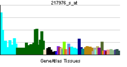
- DYNC1LI1
-
Cytoplasmic dynein 1 light intermediate chain 1 is a protein that in humans is encoded by the DYNC1LI1 gene.[1][2]
References
- ^ Pfister KK, Fisher EM, Gibbons IR, Hays TS, Holzbaur EL, McIntosh JR, Porter ME, Schroer TA, Vaughan KT, Witman GB, King SM, Vallee RB (Nov 2005). "Cytoplasmic dynein nomenclature". J Cell Biol 171 (3): 411–3. doi:10.1083/jcb.200508078. PMC 2171247. PMID 16260502. http://www.pubmedcentral.nih.gov/articlerender.fcgi?tool=pmcentrez&artid=2171247.
- ^ "Entrez Gene: DYNC1LI1 dynein, cytoplasmic 1, light intermediate chain 1". http://www.ncbi.nlm.nih.gov/sites/entrez?Db=gene&Cmd=ShowDetailView&TermToSearch=51143.
Further reading
- Maruyama K, Sugano S (1994). "Oligo-capping: a simple method to replace the cap structure of eukaryotic mRNAs with oligoribonucleotides.". Gene 138 (1-2): 171–4. doi:10.1016/0378-1119(94)90802-8. PMID 8125298.
- Suzuki Y, Yoshitomo-Nakagawa K, Maruyama K, et al. (1997). "Construction and characterization of a full length-enriched and a 5'-end-enriched cDNA library.". Gene 200 (1-2): 149–56. doi:10.1016/S0378-1119(97)00411-3. PMID 9373149.
- Tynan SH, Purohit A, Doxsey SJ, Vallee RB (2000). "Light intermediate chain 1 defines a functional subfraction of cytoplasmic dynein which binds to pericentrin.". J. Biol. Chem. 275 (42): 32763–8. doi:10.1074/jbc.M001536200. PMID 10893222.
- Bielli A, Thörnqvist PO, Hendrick AG, et al. (2001). "The small GTPase Rab4A interacts with the central region of cytoplasmic dynein light intermediate chain-1.". Biochem. Biophys. Res. Commun. 281 (5): 1141–53. doi:10.1006/bbrc.2001.4468. PMID 11243854.
- Ota T, Suzuki Y, Nishikawa T, et al. (2004). "Complete sequencing and characterization of 21,243 full-length human cDNAs.". Nat. Genet. 36 (1): 40–5. doi:10.1038/ng1285. PMID 14702039.
- Beausoleil SA, Jedrychowski M, Schwartz D, et al. (2004). "Large-scale characterization of HeLa cell nuclear phosphoproteins.". Proc. Natl. Acad. Sci. U.S.A. 101 (33): 12130–5. doi:10.1073/pnas.0404720101. PMC 514446. PMID 15302935. http://www.pubmedcentral.nih.gov/articlerender.fcgi?tool=pmcentrez&artid=514446.
- Ballif BA, Villén J, Beausoleil SA, et al. (2005). "Phosphoproteomic analysis of the developing mouse brain.". Mol. Cell Proteomics 3 (11): 1093–101. doi:10.1074/mcp.M400085-MCP200. PMID 15345747.
- Nousiainen M, Silljé HH, Sauer G, et al. (2006). "Phosphoproteome analysis of the human mitotic spindle.". Proc. Natl. Acad. Sci. U.S.A. 103 (14): 5391–6. doi:10.1073/pnas.0507066103. PMC 1459365. PMID 16565220. http://www.pubmedcentral.nih.gov/articlerender.fcgi?tool=pmcentrez&artid=1459365.
- Olsen JV, Blagoev B, Gnad F, et al. (2006). "Global, in vivo, and site-specific phosphorylation dynamics in signaling networks.". Cell 127 (3): 635–48. doi:10.1016/j.cell.2006.09.026. PMID 17081983.
Categories:- Human proteins
- Chromosome 3 gene stubs
Wikimedia Foundation. 2010.

