- Atlanto-axial joint
-
Atlanto-axial joint 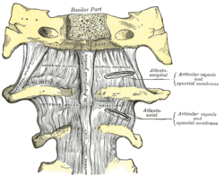
Anterior atlantoöccipital membrane and atlantoaxial ligament. 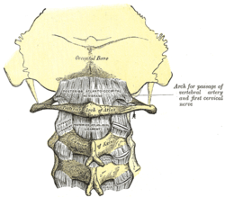
Posterior atlantoöccipital membrane and atlantoaxial ligament. Latin articulatio atlantoaxialis mediana, articulatio atlantoaxialis lateralis Gray's subject #73 292 MeSH Atlanto-Axial+Joint The Atlanto-axial joint (articulation of the atlas with the axis) is of a complicated nature. It consists of no fewer than four distinct joints.[citation needed]
There is a pivot articulation between the odontoid process of the axis and the ring formed by the anterior arch and the transverse ligament of the atlas.
Contents
Lateral and medial joints
There are two atlanto-axial joints: median and lateral:[1]
- The median atlantoaxial joint is sometimes considered a double joint:[2]
- one between the posterior surface of the anterior arch of atlas and the front of the odontoid process
- one between the anterior surface of the ligament and the back of the odontoid process
- The lateral atlantoaxial joint involves the lateral mass of atlas and axis.[3] Between the articular processes of the two bones there is on either side an arthrodial or gliding joint.
Ligaments
The ligaments connecting these bones are:
- Articular capsules
- Anterior atlantoaxial ligament
- Posterior atlantoaxial ligament
- Transverse ligament of the atlas
The atlantoaxial joint in common terminology is actually a composition of three: two lateral and one median atlantoaxial joints. Because of its proximity to the brain stem and importance in stabilization, fracture or injury at this level can be catastrophic. Common trauma and pathologies include (but are not limited to):
The Dens: significant depression on the skull can push the dens into the brainstem, causing death. The dens itself is vulnerable to fracture due to trauma or ossification.
Transverse ligament: Should the transverse ligament of the atlas fail due to trauma or disease, the dens is no longer anchored and can travel up the cervical spine, causing paralysis. If it reaches the medulla death can result. Alar ligaments: stress or trauma can stretch the weaker alar ligaments, causing an increase in range of motion of approximately 30%.
Posterior Atlanto-Occipital Membrane: genetic traits can sometimes result in ossification, turning the groove into an foramen.
Dalley, Arthur F; Moore, Keith L. Clinically Oriented Anatomy Fourth Edition. Baltimore. Lippincott Williams & Wilkins, 1992. Saladin, Kenneth S. Anatomy &Physiology: the Unity of Form and Function. New York. McGraw Hill, 2007.
Capsule
The atlantoaxial articular capsules are thin and loose, and connect the margins of the lateral masses of the atlas with those of the posterior articular surfaces of the axis.
Each is strengthened at its posterior and medial part by an accessory ligament, which is attached below to the body of the axis near the base of the odontoid process, and above to the lateral mass of the atlas near the transverse ligament.
References
- ^ Federative Committee on Anatomical Terminology (1998). Terminologia anatomica: international anatomical terminology. Thieme. pp. 27–. ISBN 9783131143617. http://books.google.com/books?id=0dYwOxPA6VcC&pg=PA27. Retrieved 17 June 2010.
- ^ Carmine D. Clemente (2010). Clemente's Anatomy Dissector. Lippincott Williams & Wilkins. pp. 361–. ISBN 9781608313846. http://books.google.com/books?id=V-M4CUkkZB0C&pg=PA361. Retrieved 17 June 2010.
- ^ ii/l/lateral_atlantoaxial_joint article at GE's Medcyclopaedia
External links
Joints and ligaments of torso (TA A03.02–04, GA 3.299) Vertebral Of vertebral bodiesAtlanto-axialmedian: Cruciate ligament of atlas (Transverse ligament of atlas) · Alar ligament · Apical ligament of dens · Tectorial membrane of atlanto-axial joint
lateral: no ligaments
anterior atlantoaxial ligament · posterior atlantoaxial ligamentno ligamentsLumbosacralThorax Radiate ligament · Intra-articular ligamentno ligamentsno ligamentsPelvis Syndesmoses of pelvic girdleanterior sacroiliac ligament · posterior sacroiliac ligament · interosseous sacroiliac ligament
ligaments connecting the sacrum and ischium: sacrotuberous ligament · sacrospinous ligament
Greater sciatic foramen · Lesser sciatic foramenM: JNT
anat(h/c, u, t, l)/phys
noco(arth/defr/back/soft)/cong, sysi/epon, injr
proc, drug(M01C, M4)
Categories:- Joints
- Vertebral column
- The median atlantoaxial joint is sometimes considered a double joint:[2]


Wikimedia Foundation. 2010.