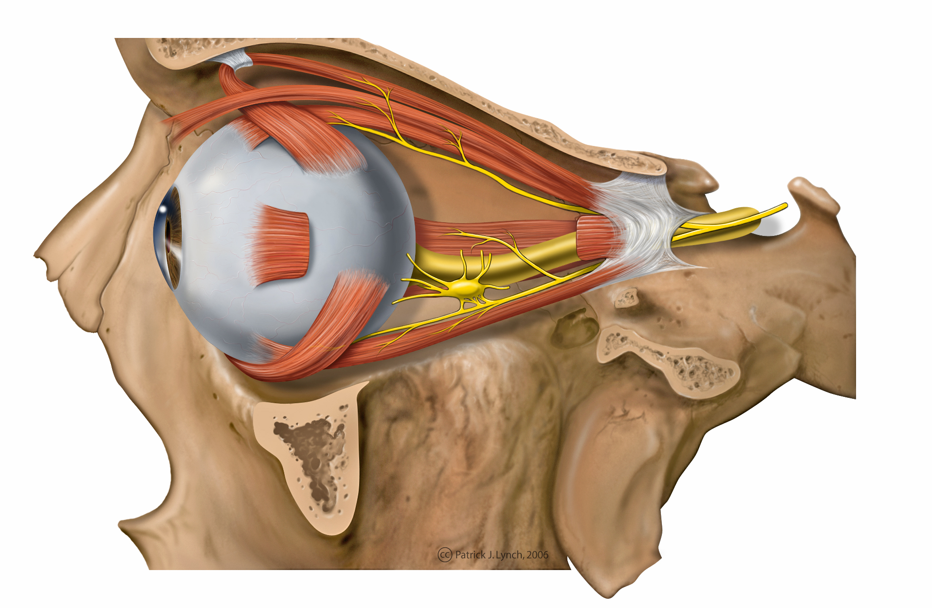
- Muscles of orbit
-
Muscle Innervation Primary function Secondary function Tertiary function Levator palpebrae superioris Oculomotor nerve Elevation of the superior (upper) eyelid . . Superior rectus Oculomotor nerve Elevation Intorsion Adduction Inferior rectus Oculomotor nerve Depression Extorsion Adduction Lateral rectus Abducens nerve Abduction . . Medial rectus Oculomotor nerve Adduction . . Superior oblique Trochlear nerve Intorsion Depression Abduction Inferior oblique Oculomotor nerve Extorsion Elevation Abduction 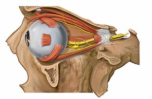 Muscles of the orbit
The muscles of the orbit are a group of six muscles that control movement of the eye. Four of the muscles control the movement of the eye in the four cardinal directions: up, down, left and right. The remaining two muscles control the adjustments involved in counteracting head movement; for instance this can be observed by looking into ones own eyes in a mirror whilst moving ones head.
A good mnemonic to remember which muscles are innervated by what nerve is to paraphrase it as a molecular equation: LR6SO4R3.
Lateral Rectus - Cranial Nerve VI,
Superior Oblique - Cranial Nerve IV,
the Rest of the muscles - Cranial Nerve III.Another way to remember which nerves innervate which muscles is to understand the meaning behind all of the Latin words. The fourth cranial nerve, the trochlear, is so named because the muscle it innervates, the superior oblique, runs through a little fascial pulley that changes its direction of pull. This pulley exists in the superiomedial corner of each orbit, and "trochl-" is Latin for "pulley." The sixth cranial nerve, the abducens, is so named because it controls the lateral rectus, which abducts the eye (rotates it laterally) upon contraction. All of the other muscles are controlled by the third cranial nerve, the oculomotor, which is so named because it is in charge of the movement (motor) of the eye (oculo-).
Contents
Purpose
The motor apparatus, precisely and rapidly, controls eye movement for exact alignment with the fovea, since this is the area that deals with sharp vision. This swift and accurate motion is evident in reading. When keeping the gaze on a small object, like a golf ball, the eyes must compensate for the small movements of the head to keep the object on the fovea. The eyes can be controlled by voluntary means, as one can deliberately change focus. However, most eye movement is done without awareness. This is evident when viewing moving objects or head or body movement. Studying the movements of the eyes depend on reflexes brought on by factors in the environment or the individual, keeping in mind the voluntary control.
Clinical Examination
The initial clinical examination of the extraoccular eye muscles is done by examining the movement of the globe of the eye through the six cardinal eye movements. When the eye is turned in (nasally) and horizontally, the function of the medial rectus muscle is being tested. When it is turned out (temporally) and horizontally, the function of the lateral rectus muscle is tested. When turning the eye down and out, the inferior rectus is contracting. Turning the eye up and out relies on the superior rectus. Paradoxically, turning the eye up and in uses the inferior oblique muscle, and turning it down and in uses the superior oblique.
All of these six movements can be tested by drawing a large "H" in the air with a finger or other object in front of a patient's face and having them follow the tip of the finger or object with their eyes without moving their head. Having them focus on the object as it is moved in toward their face in the midline will test convergence, or the eyes' ability to turn inward simultaneously to focus on a near object.
Sources
- Henry Gray: Anatomy of the human body (Bartleby.com; Great Books Online)
- Eldra Pearl Solomon; Richard R. Schmidt, Peter James Adragna (1990). Human anatomy & physiology. Saunders College Publishing. ISBN 978-0-03-011914-9. http://books.google.com/?id=N9RqAAAAMAAJ.
See also
Muscles of orbit / Extraocular muscles (TA A15.2.07, GA 10.1021) Eyelid Globe List of muscles of head and neck: the head (TA A04.1, GA 4.378) Extraocular (CN III, IV, VI) oblique (inferior, superior) · rectus (superior, inferior, medial, lateral) · levator palpebrae superioris (superior tarsal)Mastication (CN V3) masseter · temporalis (sphenomandibularis) · pterygoid (lateral, medial)
fascia: Masseteric fascia · Temporal fascia · Deep portion: cementomaxillary tendon · Superficial portion: cementomandibular tendonFacial (CN VII) levator anguli oris · levator labii superioris · zygomaticus (major, minor)
orbicularis oris · risorius · buccinator
depressor anguli oris · depressor labii inferioris · mentalisPalate/fauces (CN IX, X, XI)
(except TVP=V3)veli palatini (tensor, levator) · musculus uvulae · palatopharyngeus (to pharynx) · palatoglossus (to tongue)Tongue (CN XII) extrinsic (genioglossus, hyoglossus/chondroglossus, styloglossus, and palatoglossus) · intrinsic (superior longitudinal, inferior longitudinal, transverse, vertical)Categories:- Muscles of the head and neck
- Eye anatomy
Wikimedia Foundation. 2010.