- Hepatitis B virus
-
For the disease, see Hepatitis B.
Hepatitis B virus 
TEM micrograph showing hepatitis B viruses Virus classification Group: Group VII (dsDNA-RT) Order: Unassigned Family: Hepadnaviridae Genus: Orthohepadnavirus Species: Hepatitis B virus Hepatitis B virus, abbreviated HBV, is a species of the genus Orthohepadnavirus, which is likewise a part of the Hepadnaviridae family of viruses.[1] This virus causes the disease hepatitis B.[2]
Contents
Disease
Main article: Hepatitis BIn addition to causing hepatitis B, infection with HBV can lead to cirrhosis and hepatocellular carcinoma.[3]
It has also been suggested that it may increase the risk of pancreatic cancer.[2]
Classification
The hepatitis B virus is classified as the type species of the Orthohepadnavirus, which contains three other species: the Ground squirrel hepatitis virus, Woodchuck hepatitis virus, and the Woolly monkey hepatitis B virus. The genus is classified as part of the Hepadnaviridae family, which contains two other genera, the Avihepadnavirus and a second which has yet to be assigned. This family of viruses have not been assigned to a viral order.[4] Viruses similar to hepatitis B have been found in all the Old World apes (orangutan, gibbons, gorillas and chimpanzees) and from a New World woolly monkey suggesting an ancient origin for this virus in primates.
The virus is divided into four major serotypes (adr, adw, ayr, ayw) based on antigenic epitopes present on its envelope proteins, and into eight genotypes (A-H) according to overall nucleotide sequence variation of the genome. The genotypes have a distinct geographical distribution and are used in tracing the evolution and transmission of the virus. Differences between genotypes affect the disease severity, course and likelihood of complications, and response to treatment and possibly vaccination.[5][6]
Morphology
Structure
Hepatitis B virus is a member of the Hepadnavirus family.[7] The virus particle, (virion) consists of an outer lipid envelope and an icosahedral nucleocapsid core composed of protein. The nucleocapsid encloses the viral DNA and a DNA polymerase that has reverse transcriptase activity similar to retroviruses.[8] The outer envelope contains embedded proteins which are involved in viral binding of, and entry into, susceptible cells. The virus is one of the smallest enveloped animal viruses with a virion diameter of 42 nm, but pleomorphic forms exist, including filamentous and spherical bodies lacking a core. These particles are not infectious and are composed of the lipid and protein that forms part of the surface of the virion, which is called the surface antigen (HBsAg), and is produced in excess during the life cycle of the virus.[9]
Components
It consists of:
- HBsAg
- HBcAg (HBeAg is a splice variant)
- Hepatitis B virus DNA polymerase
- HBx. The function of this is not yet well known.[10]
Hepatitis D virus requires HBV envelope particles to become virulent.[11]
Evolution
The early evolution of the Hepatitis B, like that of all viruses, is difficult to establish.
The divergence of orthohepadnavirus and avihepadnavirus occurred ~125,000 years ago (95% interval 78,297–313,500).[12] Both the Avihepadnavirus and Orthohepadna viruses began to diversify about 25,000 years ago.[12] The branching at this time lead to the emergence of the Orthohepadna genotypes A-H. Human strains have a most recent common ancestor dating back to 7,000 (95% interval: 5,287–9,270) to 10,000 (95% interval: 6,305–16,681) years ago.
The Avihepadnavirus lack a X protein but a vestigial X reading frame is present in the genome of duck hepadnavirus.[13] The X protein may have evolved from a DNA glycosylase.
The rate of nonsynonymous mutations in this virus has been estimated to be about 2×10⁻⁵ amino acid replacements per site per year.[14] The mean number of nucleotide substitutions/site/year is ~7.9 x 10⁻⁵.
Genome
 The genome organisation of HBV. The genes overlap.
Size
The genome of HBV is made of circular DNA, but it is unusual because the DNA is not fully double-stranded. One end of the full length strand is linked to the viral DNA polymerase. The genome is 3020-3320 nucleotides long (for the full length strand) and 1700-2800 nucleotides long (for the short length strand).[15]
Encoding
The negative-sense, (non-coding), is complementary to the viral mRNA. The viral DNA is found in the nucleus soon after infection of the cell. The partially double-stranded DNA is rendered fully double-stranded by completion of the (+) sense strand and removal of a protein molecule from the (-) sense strand and a short sequence of RNA from the (+) sense strand. Non-coding bases are removed from the ends of the (-)sense strand and the ends are rejoined.
There are four known genes encoded by the genome called C, X, P, and S. The core protein is coded for by gene C (HBcAg), and its start codon is preceded by an upstream in-frame AUG start codon from which the pre-core protein is produced. HBeAg is produced by proteolytic processing of the pre-core protein. The DNA polymerase is encoded by gene P. Gene S is the gene that codes for the surface antigen (HBsAg). The HBsAg gene is one long open reading frame but contains three in frame "start" (ATG) codons that divide the gene into three sections, pre-S1, pre-S2, and S. Because of the multiple start codons, polypeptides of three different sizes called large, middle, and small (pre-S1 + pre-S2 + S, pre-S2 + S, or S) are produced.[16] The function of the protein coded for by gene X is not fully understood.[17]
Several non-coding RNA elements have been identified in the HBV genome. These include: HBV PREalpha, HBV PREbeta and HBV RNA encapsidation signal epsilon.[18][19]
Genotypes
There are eight known genotypes labeled A through H.[5] A possible new "I" genotype has been described,[20] but acceptance of this notation is not universal.[21] Different genotypes may respond to treatment in different ways.[22][23]
The genotypes differ by at least 8% of the sequence and have distinct geographical distributions and this has been associated with anthropological history. Type F which diverges from the other genomes by 14% is the most divergent type known. Type A is prevalent in Europe, Africa and South-east Asia, including the Philippines. Type B and C are predominant in Asia; type D is common in the Mediterranean area, the Middle East and India; type E is localized in sub-Saharan Africa; type F (or H) is restricted to Central and South America. Type G has been found in France and Germany. Genotypes A, D and F are predominant in Brazil and all genotypes occur in the United States with frequencies dependent on ethnicity.
The E and F strains appear to have originated in aboriginal populations of Africa and the New World, respectively.
Within genotypes 24 subtypes have been described which differ by 4-8% of the genome.
Type A has two subtypes: Aa (A1) in Africa/Asia and the Philippines and Ae (A2) in Europe/United States.
Type B has two distinct geographical distributions: Bj/B1 ('j' - Japan) and Ba/B2 ('a' - Asia). Type Ba has been further subdivided into four clades (B2 - B4).
Type C has two geographically subtypes: Cs (C1) in South-east Asia and Ce (C2) in East Asia. The C subtypes have been divided into five clades (C1 - C5). A sixth clade (C6) has been described in the Philippines but only in one isolate to date.[24] Type C1 is associated with Vietnam, Myanmar and Thailand; type C2 with Japan, Korea and China; type C3 with New Caledonia and Polynesia; C4 with Australia; and C5 with the Philippines. A further subtype has been described in Papua, Indonesia.[25]
Type D has been divided into 7 subtypes (D1 - D7).
Type F has been subdivided into 4 subtypes (F1 - F4). F1 has been further divided in to 1a and 1b. In Venezuela subtypes F1, F2, and F3 are found in East and West Amerindians. Among South Amerindians only F3 was found. Subtypes Ia, III, and IV exhibit a restricted geographic distribution (Central America, the North and the South of South America respectively) while clades Ib and II are found in all the Americas except in the Northern South America and North America respectively.
Life cycle
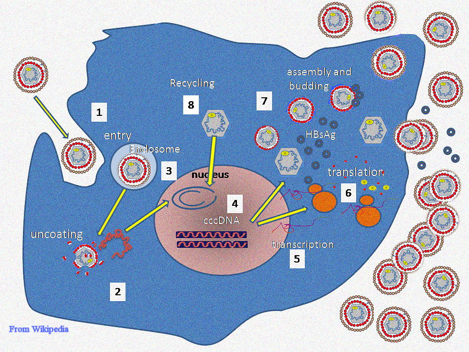
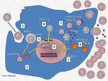 Hepatitis B virus replication .
The life cycle of hepatitis B virus is complex. Hepatitis B is one of a few known non-retroviral viruses which use reverse transcription as a part of its replication process.
- Attachment
- The virus gains entry into the cell by binding to a receptor on the surface of the cell and enters it by endocytosis. The cell surface receptor has yet to be identified however it is suspected to be a member of the ovalbumin family of serine protease inhibitors.[1]
- Penetration
- The virus membrane then fuses with the host cell's membrane releasing the DNA and core proteins into the cytoplasm.
- Uncoating
- Because the virus multiplies via RNA made by a host enzyme, the viral genomic DNA has to be transferred to the cell nucleus by host proteins called chaperones. The core proteins dissociate from the partially double stranded viral DNA is then made fully double stranded and transformed into covalently closed circular DNA (cccDNA) that serves as a template for transcription of four viral mRNAs.
- Replication
- The largest mRNA, (which is longer than the viral genome), is used to make the new copies of the genome and to make the capsid core protein and the viral DNA polymerase.
- Assembly
- These four viral transcripts undergo additional processing and go on to form progeny virions which are released from the cell or returned to the nucleus and re-cycled to produce even more copies.[16][26]
- Release
- The long mRNA is then transported back to the cytoplasm where the virion P protein synthesizes DNA via its reverse transcriptase activity.
See also
References
- ^ a b Hunt, Richard (2007-11-21). "Hepatitis viruses". University of Southern California, Department of Pathology and Microbiology. http://pathmicro.med.sc.edu/virol/hepatitis-virus.htm. Retrieved 2008-03-13.
- ^ a b Hassan MM, Li D, El-Deeb AS, et al. (October 2008). "Association between hepatitis B virus and pancreatic cancer". J. Clin. Oncol. 26 (28): 4557–62. doi:10.1200/JCO.2008.17.3526. PMC 2562875. PMID 18824707. http://www.jco.org/cgi/pmidlookup?view=long&pmid=18824707.
- ^ Schwalbe M, Ohlenschläger O, Marchanka A, et al. (March 2008). "Solution structure of stem-loop alpha of the hepatitis B virus post-transcriptional regulatory element". Nucleic Acids Res. 36 (5): 1681–9. doi:10.1093/nar/gkn006. PMC 2275152. PMID 18263618. http://nar.oxfordjournals.org/cgi/pmidlookup?view=long&pmid=18263618.
- ^ "00.030. Hepadnaviridae - ICTVdB Index of Viruses". International Committee on Taxonomy of Viruses. 2008-07-08. http://www.ncbi.nlm.nih.gov/ICTVdb/Ictv/fs_hepad.htm. Retrieved 2009-03-13.
- ^ a b Kramvis A, Kew M, François G (2005). "Hepatitis B virus genotypes". Vaccine 23 (19): 2409–23. doi:10.1016/j.vaccine.2004.10.045. PMID 15752827.
- ^ Magnius LO, Norder H (1995). "Subtypes, genotypes and molecular epidemiology of the hepatitis B virus as reflected by sequence variability of the S-gene". Intervirology 38 (1–2): 24–34. PMID 8666521.
- ^ Zuckerman AJ (1996). Hepatitis Viruses. In: Baron's Medical Microbiology (Baron S et al., eds.) (4th ed.). Univ of Texas Medical Branch. ISBN 0-9631172-1-1. http://www.ncbi.nlm.nih.gov/books/bv.fcgi?rid=mmed.section.3738.
- ^ Locarnini S (2004). "Molecular virology of hepatitis B virus". Semin. Liver Dis. 24 Suppl 1: 3–10. doi:10.1055/s-2004-828672. PMID 15192795.
- ^ Howard CR (1986). "The biology of hepadnaviruses". J. Gen. Virol. 67 ( Pt 7) (7): 1215–35. doi:10.1099/0022-1317-67-7-1215. PMID 3014045.
- ^ Guo GH, Tan DM, Zhu PA, Liu F (February 2009). "Hepatitis B virus X protein promotes proliferation and upregulates TGF-beta1 and CTGF in human hepatic stellate cell line, LX-2". Hbpd Int 8 (1): 59–64. PMID 19208517. http://www.hbpdint.com/text.asp?id=1196.
- ^ Chai N, Chang HE, Nicolas E, Han Z, Jarnik M, Taylor J (August 2008). "Properties of subviral particles of hepatitis B virus". J. Virol. 82 (16): 7812–7. doi:10.1128/JVI.00561-08. PMC 2519590. PMID 18524834. http://jvi.asm.org/cgi/pmidlookup?view=long&pmid=18524834.
- ^ a b van Hemert FJ, van de Klundert MA, Lukashov VV, Kootstra NA, Berkhout B, Zaaijer HL (2011) Protein x of hepatitis B virus: origin and structure similarity with the central domain of DNA glycosylase. PLoS One. 2011;6(8):e23392.
- ^ Lin B, Anderson DA. A vestigial X open reading frame in duck hepatitis B virus. Intervirol. 2000;43:185–190.
- ^ Osiowy C, Giles E, Tanaka Y, Mizokami M, Minuk GY (2006). "Molecular evolution of hepatitis B virus over 25 years". J Virol 80 (21): 10307–10314.
- ^ Kay A, Zoulim F (2007). "Hepatitis B virus genetic variability and evolution". Virus Res. 127 (2): 164–76. doi:10.1016/j.virusres.2007.02.021. PMID 17383765.
- ^ a b Beck J, Nassal M (2007). "Hepatitis B virus replication". World J. Gastroenterol. 13 (1): 48–64. PMID 17206754.
- ^ Bouchard MJ, Schneider RJ (2004). "The enigmatic X gene of hepatitis B virus". J. Virol. 78 (23): 12725–34. doi:10.1128/JVI.78.23.12725-12734.2004. PMC 524990. PMID 15542625. http://www.pubmedcentral.nih.gov/articlerender.fcgi?tool=pmcentrez&artid=524990.
- ^ Smith Gj, 3rd; Donello, JE; Lück, R; Steger, G; Hope, TJ (1998). "The hepatitis B virus post-transcriptional regulatory element contains two conserved RNA stem-loops which are required for function". Nucleic Acids Research 26 (21): 4818–27. doi:10.1093/nar/26.21.4818. PMC 147918. PMID 9776740. http://www.pubmedcentral.nih.gov/articlerender.fcgi?tool=pmcentrez&artid=147918.
- ^ Flodell S, Schleucher J, Cromsigt J, Ippel H, Kidd-Ljunggren K, Wijmenga S (November 2002). "The apical stem-loop of the hepatitis B virus encapsidation signal folds into a stable tri-loop with two underlying pyrimidine bulges". Nucleic Acids Res. 30 (21): 4803–11. doi:10.1093/nar/gkf603. PMC 135823. PMID 12409471. http://nar.oxfordjournals.org/cgi/pmidlookup?view=long&pmid=12409471.
- ^ Olinger CM, Jutavijittum P, Hübschen JM, et al. (November 2008). "Possible new hepatitis B virus genotype, southeast Asia". Emerging Infect. Dis. 14 (11): 1777–80. doi:10.3201/eid1411.080437. PMC 2630741. PMID 18976569. http://www.cdc.gov/eid/content/14/11/1777.htm.
- ^ Kurbanov F, Tanaka Y, Kramvis A, Simmonds P, Mizokami M (August 2008). "When should "I" consider a new hepatitis B virus genotype?". J. Virol. 82 (16): 8241–2. doi:10.1128/JVI.00793-08. PMC 2519592. PMID 18663008. http://jvi.asm.org/cgi/pmidlookup?view=long&pmid=18663008.
- ^ Palumbo E (2007). "Hepatitis B genotypes and response to antiviral therapy: a review". Am J Ther 14 (3): 306–9. doi:10.1097/01.pap.0000249927.67907.eb. PMID 17515708. http://meta.wkhealth.com/pt/pt-core/template-journal/lwwgateway/media/landingpage.htm?an=00045391-200705000-00016.
- ^ Mahtab MA, Rahman S, Khan M, Karim F (October 2008). "Hepatitis B virus genotypes: an overview". Hbpd Int 7 (5): 457–64. PMID 18842489. http://www.hbpdint.com/text.asp?id=1142.
- ^ Cavinta L., Sun J., May A., Yin J., von Meltzer M., Radtke M., Barzaga N.G., Cao G., Schaefer S. et al. (2009). "A new isolate of hepatitis B virus from the Philippines possibly representing a new subgenotype C6". J. Med. Virol 81 (6): 983–987.
- ^ Lusida M.I., Nugrahaputra V.E., Soetjipto Handajani R., Nagano-Fujii M., Sasayama M., Utsumi T., Hotta H. (2008). "Novel subgenotypes of hepatitis B virus genotypes C and D in Papua, Indonesia". J. Clin. Microbiol 46 (7): 2160–2166. doi:10.1128/JCM.01681-07. PMC 2446895. PMID 18463220. http://www.pubmedcentral.nih.gov/articlerender.fcgi?tool=pmcentrez&artid=2446895.
- ^ Bruss V (2007). "Hepatitis B virus morphogenesis". World J. Gastroenterol. 13 (1): 65–73. PMID 17206755.
Infectious diseases · Viral systemic diseases (A80–B34, 042–079) Oncovirus Immune disorders Central
nervous systemEncephalitis/
meningitisDNA virus: JCV (Progressive multifocal leukoencephalopathy)
RNA virus: MeV (Subacute sclerosing panencephalitis) · LCV (Lymphocytic choriomeningitis) · Arbovirus encephalitis · Orthomyxoviridae (probable) (Encephalitis lethargica) · RV (Rabies) · Chandipura virus · Herpesviral meningitis · Ramsay Hunt syndrome type IIEyeCardiovascular Respiratory system/
acute viral nasopharyngitis/
viral pneumoniaDigestive system Urogenital DNA Hepatitis BRNA capsid: matrix protein (M1 protein) · viral envelope (M2 protein)
glycoprotein: Influenza hemagglutinin · NeuraminidaseParainfluenzaParainfluenza hemagglutinin-neuraminidaseRSVRespiratory syncytial virus G proteinRT VSPs: gag · pol (Integrase, Reverse transcriptase, HIV-1 protease) · env (gp120, gp41)
VRAPs: transactivators (Tat, Rev, Vpr) · Nef · Vif · VpuFusion protein Categories:- Hepatitis
- Pararetroviruses


Wikimedia Foundation. 2010.