- Electron crystallography
-
Electron crystallography is a method to determine the arrangement of atoms in solids using a transmission electron microscope (TEM).
Contents
Comparison with X-ray crystallography
It can complement X-ray crystallography for studies of very small crystals (<0.1 micrometers), both inorganic, organic and proteins, such as membrane proteins, that cannot easily form the large 3-dimensional crystals required for that process. Protein structures are usually determined from either 2-dimensional crystals (sheets or helices), polyhedrons such as viral capsids, or dispersed individual proteins. Electrons can be used in these situations, whereas X-rays cannot, because electrons interact more strongly with atoms than X-rays do. Thus, X-rays will travel through a thin 2-dimensional crystal without diffracting significantly, whereas electrons can be used to form an image. Conversely, the strong interaction between electrons and proteins makes thick (e.g. 3-dimensional > 1 micrometer) crystals impervious to electrons, which only penetrate short distances.
One of the main difficulties in X-ray crystallography is determining phases in the diffraction pattern. Because no X-ray lens exists, X-rays cannot be used to form an image of the crystal being diffracted, and hence phase information is lost. Fortunately, electron microscopes contain electron lenses, and so the crystallographic structure factor phase information can be experimentally determined in electron crystallography. Aaron Klug was the first to realise that the phase information could be read out directly from the Fourier transform of an electron microscopy image that had been scanned into a computer, already in 1968. For this, and his studies on virus structures and transfer-RNA, Klug received the Nobel Prize for chemistry in 1982.
Radiation damage
A common problem to X-ray crystallography and electron crystallography is radiation damage, by which especially organic molecules and proteins are damaged as they are being imaged, limiting the resolution that can be obtained. This is especially troublesome in the setting of electron crystallography, where that radiation damage is focused on far fewer atoms. One technique used to limit radiation damage is electron cryomicroscopy, in which the samples undergo cryofixation and imaging takes place at liquid nitrogen or even liquid helium temperatures. Because of this problem, X-ray crystallography has been much more successful in determining the structure of proteins that are especially vulnerable to radiation damage.
Protein structures determined by electron crystallography
The first electron crystallographic protein structure to achieve atomic resolution was bacteriorhodopsin, determined by Richard Henderson and coworkers at the Medical Research Council Laboratory of Molecular Biology in 1990. However, already in 1975 Unwin and Henderson had determined the first membrane protein structure at intermediate resolution (7 Ångström), showing for the first time the internal structure of a membrane protein, with its alpha-helices standing perpendicular to the plane of the membrane. Since then, several other high-resolution structures have been determined by electron crystallography, including the light-harvesting complex,[1] the nicotinic acetylcholine receptor,[2] and the bacterial flagellum.[3]
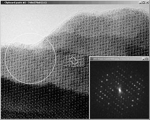 Electron microscopy image of an inorganic tantalum oxide, with its Fourier transform, also called diffractogram, inset. Notice how the appearance changes from the upper thin region to the thicker lower region. The unit cell of this compound is about 15 by 25 Ångström. It is outlined at the centre of the figure, inside the result from image processing, where the symmetry has been taken into account. The black dots show clearly all the tantalum atoms. The diffraction extends to 6 orders along the 15 Å direction and 10 orders in the perpendicular direction. Thus the resolution of the EM image is 2.5 Å (15/6 or 25/10). This calculated Fourier transform contain both amplitudes (as seen) and phases (not displayed).
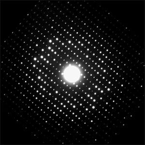 Electron diffraction pattern of the same crystal of inorganic tantalum oxide shown above. Notice that there are many more diffraction spots here than in the diffractogram calculated from the EM image above. The diffraction extends to 12 orders along the 15 Å direction and 20 orders in the perpendicular direction. Thus the resolution of the ED pattern is 1.25 Å (15/12 or 25/20). ED patterns do not contain phase information, but the clear differences between intensities of the diffraction spots can be used in crystal structure determination.
Application to inorganic materials
Electron crystallographic studies on inorganic crystals using high-resolution electron microscopy (HREM) images were first performed by Aaron Klug in 1978[4] and by Sven Hovmöller and coworkers in 1984.[5] HREM images were used because they allow to select (by computer software) only the very thin regions close to the edge of the crystal for structure analysis (see also crystallographic image processing). This is of crucial importance since in the thicker parts of the crystal the exit-wave function (which carries the information about the intensity and position of the projected atom columns) is no longer linearly related to the projected crystal structure. Moreover not only do the HREM images change their appearance with increasing crystal thickness, they are also very sensitive to the chosen setting of the defocus Δf of the objective lens (see the HREM images of GaN for example). To cope with this complexity Michael O'Keefe started in the early 1970s to develop image simulation software which allowed to understand an interpret the observed contrast changes in HREM images.[6]
There was a serious disagreement in the field of electron microscopy of inorganic compounds; while some have claimed that "the phase information is present in EM images" others have the opposite view that "the phase information is lost in EM images". The reason for these opposite views is that the word "phase" has been used with different meanings in the two communities of physicists and crystallographers. The physicists are more concerned about the "electron wave phase" - the phase of a wave moving through the sample during exposure by the electrons. This wave has a wavelength of about 0.02-0.03 Ångström (depending on the accelerating voltage of the electron microscope). Its phase is related to the phase of the undiffracted direct electron beam. The crystallographers, on the other hand, mean the "crystallographic structure factor phase" when they simply say "phase". This phase is the phase of standing waves of potential in the crystal (very similar to the electron density measured in X-ray crystallography). Each of these waves have their specific wavelength, called d-value for distance between so-called Bragg planes of low/high potential. These d-values range from the unit cell dimensions to the resolution limit of the electron microscope, i.e. typically from 10 or 20 Ångströms down to 1 or 2 Ångströms. Their phases are related to a fixed point in the crystal, defined in relation to the symmetry elements of that crystal. The crystallographic phases are a property of the crystal, so they exist also outside the electron microscope. The electron waves vanish if the microscope is switched off. In order to determine a crystal structure, it is necessary to know the crystallographic structure factors, but not to know the electron wave phases. A more detailed discussion how (crystallographic structure factor) phases link with the phases of the electron wave can be found in.[7]
Just as with proteins, it has been possible to determine the atomic structures of inorganic crystals by electron crystallography. For simpler structure it is sufficient to use three perpendicular views, but for more complicated structures, also projections down ten or more different diagonals may be needed.
In addition to electron microscopy images, it is also possible to use electron diffraction (ED) patterns for crystal structure determination.[8][9] The utmost care must be taken to record such ED patterns from the thinnest areas in order to keep most of the structure related intensity differences between the reflections (quasi-kinematical diffraction conditions). Just as with X-ray diffraction patterns, the important crystallographic structure factor phases are lost in electron diffraction patterns and must be uncovered by special crystallographic methods such as direct methods, maximum likelihood or (more recently) by the charge-flipping method. On the other hand, ED patterns of inorganic crystals have often a high resolution (= interplanar spacings with high Miller indices) much below 1 Ångström. This is comparable to the point resolution of the best electron microscopes. Under favourable conditions it is possible to use ED patterns from a single orientation to determine the complete crystal structure.[10] Alternatively a hybrid approach can be used which uses HRTEM images for solving and intensities from ED for refining the crystal structure.[11][12]
Recent progress for structure analysis by ED was made by introducing the Vincent-Midgley precession technique for recording electron diffraction patterns.[13] The thereby obtained intensities are usually much closer to the kinematical intensities, so that even structures can be determined that are out of range when processing conventional (selected area) electron diffraction data.[14][15]
Crystal structures determined via electron crystallography can be checked for their quality by using first-principles calculations within density functional theory (DFT). This approach was for the first time applied for the validation of several metal-rich structures which were only accessible by HRTEM and ED, respectively.[16][17]
Recently, two very complicated zeolite structures have been determined by electron crystallography combined with X-ray powder diffraction.[18][19] These are more complex than the most complex zeolite structures determined by X-ray crystallography.
References
- ^ PMID 8107845
- ^ PMID 12827192
- ^ PMID 12904785
- ^ Klug, A (1978/79) Image Analysis and Reconstruction in the Electron Microscopy of Biological Macromolecules Chemica Scripta vol 14, p. 245-256.
- ^ Hovmöller, Sven; Sjögren, Agneta; Farrants, George; Sundberg, Margareta; Marinder, Bengt-Olov (1984). "Accurate atomic positions from electron microscopy". Nature 311 (5983): 238. Bibcode 1984Natur.311..238H. doi:10.1038/311238a0.
- ^ O'Keefe, M. A.; Buseck, P. R.; Iijima, S. (1978). "Computed crystal structure images for high resolution electron microscopy". Nature 274 (5669): 322. Bibcode 1978Natur.274..322O. doi:10.1038/274322a0.
- ^ Zou, X (1999). "On the phase problem in electron microscopy: the relationship between structure factors, exit waves, and HREM images.". Microscopy research and technique 46 (3): 202–19. doi:10.1002/(SICI)1097-0029(19990801)46:3<202::AID-JEMT4>3.0.CO;2-8. PMID 10420175.
- ^ B. K. Vainshtein (1964), Structure Analysis by Electron Diffraction, Pergamon Press Oxford
- ^ D. L. Dorset (1995), Structural Electron Crystallography, Plenum Publishing Corporation ISBN 0-306-45049-6
- ^ Weirich, TE; Zou, X; Ramlau, R; Simon, A; Cascarano, GL; Giacovazzo, C; Hovmöller, S (2000). "Structures of nanometre-size crystals determined from selected-area electron diffraction data.". Acta Crystallographica A 56 (Pt 1): 29–35. doi:10.1107/S0108767399009605. PMID 10874414.
- ^ Zandbergen, H. W. (1997). "Structure Determination of Mg5Si6 Particles in Al by Dynamic Electron Diffraction Studies". Science 277 (5330): 1221. doi:10.1126/science.277.5330.1221.
- ^ Weirich, Thomas E.; Ramlau, Reiner; Simon, Arndt; Hovmöller, Sven; Zou, Xiaodong (1996). "A crystal structure determined with 0.02 Å accuracy by electron microscopy". Nature 382 (6587): 144. Bibcode 1996Natur.382..144W. doi:10.1038/382144a0.
- ^ Precession Electron Diffraction
- ^ Gemmi, M; Zou, X; Hovmöller, S; Migliori, A; Vennström, M; Andersson, Y (2003). "Structure of Ti2P solved by three-dimensional electron diffraction data collected with the precession technique and high-resolution electron microscopy.". Acta Crystallographica 59 (Pt 2): 117–26. doi:10.1107/S0108767302022559. PMID 12604849.
- ^ Weirich, T; Portillo, J; Cox, G; Hibst, H; Nicolopoulos, S (2006). "Ab initio determination of the framework structure of the heavy-metal oxide CsxNb2.54W2.46O14 from 100kV precession electron diffraction data". Ultramicroscopy 106 (3): 164–75. doi:10.1016/j.ultramic.2005.07.002. PMID 16137828.
- ^ Albe, K; Weirich, TE (2003). "Structure and stability of alpha- and beta-Ti2Se. Electron diffraction versus density-functional theory calculations.". Acta Crystallographica A 59 (Pt 1): 18–21. doi:10.1107/S0108767302018275. PMID 12496457.
- ^ Weirich, TE (2004). "First-principles calculations as a tool for structure validation in electron crystallography.". Acta Crystallographica A 60 (Pt 1): 75–81. doi:10.1107/S0108767303025042. PMID 14691330.
- ^ Gramm, Fabian; Baerlocher, Christian; McCusker, Lynne B.; Warrender, Stewart J.; Wright, Paul A.; Han, Bada; Hong, Suk Bong; Liu, Zheng et al. (2006). "Complex zeolite structure solved by combining powder diffraction and electron microscopy". Nature 444 (7115): 79–81. Bibcode 2006Natur.444...79G. doi:10.1038/nature05200. PMID 17080087.
- ^ Baerlocher, C.; Gramm, F.; Massuger, L.; McCusker, L. B.; He, Z.; Hovmoller, S.; Zou, X. (2007). "Structure of the Polycrystalline Zeolite Catalyst IM-5 Solved by Enhanced Charge Flipping". Science 315 (5815): 1113–6. Bibcode 2007Sci...315.1113B. doi:10.1126/science.1137920. PMID 17322057.
Further reading
- Zou, XD, Hovmöller, S. and Oleynikov, P. "Electron Crystallography - Electron microscopy and Electron Diffraction". IUCr Texts on Crystallography 16, Oxford university press 2011. http://ukcatalogue.oup.com/product/9780199580200.do ISBN 978-0-19-958020-0
- K. H. Downing, H. Meisheng, H.-R. Wenk & M. A. O'Keefe (1990). "Resolution of oxygen atoms in staurolite by three-dimensional transmission electron microscopy." Nature 348, p. 525 - 528.
- Zou, X.D. and Hovmöller, S. (2008). "Electron crystallography: Imaging and Single Crystal Diffraction from Powders." Acta Crystallographica A 64, p. 149-160.
- T.E. Weirich, X.D. Zou & J.L. Lábár (2006). Electron Crystallography: Novel Approaches for Structure Determination of Nanosized Materials. Springer Netherlands, ISBN 978-1402039195
External links
- Interview with Aaron Klug Nobel Laureate for work on crystallograph electron microscopy Freeview video by the Vega Science Trust.
- Raunser, S; Walz, T (2009). "Electron Crystallography as a Technique to Study the Structure on Membrane Proteins in a Lipidic Environment". Annual Review of Biophysics 38 (1): 89–105. doi:10.1146/annurev.biophys.050708.133649. PMID 19416061. http://arjournals.annualreviews.org/doi/abs/10.1146/annurev.biophys.050708.133649.
Protein structural analysis High resolution Medium resolution Fiber diffraction | Mass spectrometry | SAXSSpectroscopic Translational Diffusion Rotational Diffusion Chemical Thermodynamic Computational ←Tertiary structureCategories:


Wikimedia Foundation. 2010.