- DYNLL1
-
Dynein light chain 1, cytoplasmic is a protein that in humans is encoded by the DYNLL1 gene.[1][2][3][4]
Cytoplasmic dyneins are large enzyme complexes with a molecular mass of about 1,200 kD. They contain two force-producing heads formed primarily from dynein heavy chains, and stalks linking the heads to a basal domain, which contains a varying number of accessory intermediate chains. The complex is involved in intracellular transport and motility. The protein described in this record is a light chain and exists as part of this complex but also physically interacts with and inhibits the activity of neuronal nitric oxide synthase. Binding of this protein destabilizes the neuronal nitric oxide synthase dimer, a conformation necessary for activity, and it may regulate numerous biologic processes through its effects on nitric oxide synthase activity. Alternate transcriptional splice variants have been characterized.[4]
Interactions
DYNLL1 has been shown to interact with DLGAP1,[5] MYO5A,[5] DYNC1I1,[6] BCL2L11,[7][8] NRF1,[9] IκBα,[10] PAK1,[8] DLG4[5] and TP53BP1.[11]
References
- ^ Dick T, Ray K, Salz HK, Chia W (Jun 1996). "Cytoplasmic dynein (ddlc1) mutations cause morphogenetic defects and apoptotic cell death in Drosophila melanogaster". Mol Cell Biol 16 (5): 1966–77. PMC 231184. PMID 8628263. http://www.pubmedcentral.nih.gov/articlerender.fcgi?tool=pmcentrez&artid=231184.
- ^ Jaffrey SR, Snyder SH (Dec 1996). "PIN: an associated protein inhibitor of neuronal nitric oxide synthase". Science 274 (5288): 774–7. doi:10.1126/science.274.5288.774. PMID 8864115.
- ^ Pfister KK, Fisher EM, Gibbons IR, Hays TS, Holzbaur EL, McIntosh JR, Porter ME, Schroer TA, Vaughan KT, Witman GB, King SM, Vallee RB (Nov 2005). "Cytoplasmic dynein nomenclature". J Cell Biol 171 (3): 411–3. doi:10.1083/jcb.200508078. PMC 2171247. PMID 16260502. http://www.pubmedcentral.nih.gov/articlerender.fcgi?tool=pmcentrez&artid=2171247.
- ^ a b "Entrez Gene: DYNLL1 dynein, light chain, LC8-type 1". http://www.ncbi.nlm.nih.gov/sites/entrez?Db=gene&Cmd=ShowDetailView&TermToSearch=8655.
- ^ a b c Naisbitt, S; Valtschanoff J, Allison D W, Sala C, Kim E, Craig A M, Weinberg R J, Sheng M (Jun. 2000). "Interaction of the postsynaptic density-95/guanylate kinase domain-associated protein complex with a light chain of myosin-V and dynein". J. Neurosci. (UNITED STATES) 20 (12): 4524–34. ISSN 0270-6474. PMID 10844022.
- ^ Diefenbach, Russell J; Diefenbach Eve, Douglas Mark W, Cunningham Anthony L (Dec. 2002). "The heavy chain of conventional kinesin interacts with the SNARE proteins SNAP25 and SNAP23". Biochemistry (United States) 41 (50): 14906–15. doi:10.1021/bi026417u. ISSN 0006-2960. PMID 12475239.
- ^ Day, Catherine L; Puthalakath Hamsa, Skea Gretchen, Strasser Andreas, Barsukov Igor, Lian Lu-Yun, Huang David C S, Hinds Mark G (Feb. 2004). "Localization of dynein light chains 1 and 2 and their pro-apoptotic ligands". Biochem. J. (England) 377 (Pt 3): 597–605. doi:10.1042/BJ20031251. PMC 1223895. PMID 14561217. http://www.pubmedcentral.nih.gov/articlerender.fcgi?tool=pmcentrez&artid=1223895.
- ^ a b Vadlamudi, Ratna K; Bagheri-Yarmand Rozita, Yang Zhibo, Balasenthil Seetharaman, Nguyen Diep, Sahin Aysegul A, den Hollander Petra, Kumar Rakesh (Jun. 2004). "Dynein light chain 1, a p21-activated kinase 1-interacting substrate, promotes cancerous phenotypes". Cancer Cell (United States) 5 (6): 575–85. doi:10.1016/j.ccr.2004.05.022. ISSN 1535-6108. PMID 15193260.
- ^ Herzig, R P; Andersson U, Scarpulla R C (Dec. 2000). "Dynein light chain interacts with NRF-1 and EWG, structurally and functionally related transcription factors from humans and drosophila". J. Cell. Sci. (ENGLAND) 113 Pt 23: 4263–73. ISSN 0021-9533. PMID 11069771.
- ^ Crépieux, P; Kwon H, Leclerc N, Spencer W, Richard S, Lin R, Hiscott J (Dec. 1997). "I kappaB alpha physically interacts with a cytoskeleton-associated protein through its signal response domain". Mol. Cell. Biol. (UNITED STATES) 17 (12): 7375–85. ISSN 0270-7306. PMC 232593. PMID 9372968. http://www.pubmedcentral.nih.gov/articlerender.fcgi?tool=pmcentrez&artid=232593.
- ^ Lo, Kevin W-H; Kan Ho-Man, Chan Ling-Nga, Xu Wei-Guang, Wang Ke-Peng, Wu Zhenguo, Sheng Morgan, Zhang Mingjie (Mar. 2005). "The 8-kDa dynein light chain binds to p53-binding protein 1 and mediates DNA damage-induced p53 nuclear accumulation". J. Biol. Chem. (United States) 280 (9): 8172–9. doi:10.1074/jbc.M411408200. ISSN 0021-9258. PMID 15611139.
Further reading
- Robertson NG, Khetarpal U, Gutiérrez-Espeleta GA, et al. (1995). "Isolation of novel and known genes from a human fetal cochlear cDNA library using subtractive hybridization and differential screening". Genomics 23 (1): 42–50. doi:10.1006/geno.1994.1457. PMID 7829101.
- Crépieux P, Kwon H, Leclerc N, et al. (1997). "I kappaB alpha physically interacts with a cytoskeleton-associated protein through its signal response domain". Mol. Cell. Biol. 17 (12): 7375–85. PMC 232593. PMID 9372968. http://www.pubmedcentral.nih.gov/articlerender.fcgi?tool=pmcentrez&artid=232593.
- Tochio H, Ohki S, Zhang Q, et al. (1998). "Solution structure of a protein inhibitor of neuronal nitric oxide synthase". Nat. Struct. Biol. 5 (11): 965–9. doi:10.1038/2940. PMID 9808041.
- Rodríguez-Crespo I, Straub W, Gavilanes F, Ortiz de Montellano PR (1998). "Binding of dynein light chain (PIN) to neuronal nitric oxide synthase in the absence of inhibition". Arch. Biochem. Biophys. 359 (2): 297–304. doi:10.1006/abbi.1998.0928. PMID 9808772.
- Fan JS, Zhang Q, Li M, et al. (1999). "Protein inhibitor of neuronal nitric-oxide synthase, PIN, binds to a 17-amino acid residue fragment of the enzyme". J. Biol. Chem. 273 (50): 33472–81. doi:10.1074/jbc.273.50.33472. PMID 9837926.
- Puthalakath H, Huang DC, O'Reilly LA, et al. (1999). "The proapoptotic activity of the Bcl-2 family member Bim is regulated by interaction with the dynein motor complex". Mol. Cell 3 (3): 287–96. doi:10.1016/S1097-2765(00)80456-6. PMID 10198631.
- Liang J, Jaffrey SR, Guo W, et al. (1999). "Structure of the PIN/LC8 dimer with a bound peptide". Nat. Struct. Biol. 6 (8): 735–40. doi:10.1038/11501. PMID 10426949.
- Epstein E, Sela-Brown A, Ringel I, et al. (2000). "Dynein light chain binding to a 3′-untranslated sequence mediates parathyroid hormone mRNA association with microtubules". J. Clin. Invest. 105 (4): 505–12. doi:10.1172/JCI8557. PMC 289163. PMID 10683380. http://www.pubmedcentral.nih.gov/articlerender.fcgi?tool=pmcentrez&artid=289163.
- Naisbitt S, Valtschanoff J, Allison DW, et al. (2000). "Interaction of the postsynaptic density-95/guanylate kinase domain-associated protein complex with a light chain of myosin-V and dynein". J. Neurosci. 20 (12): 4524–34. PMID 10844022.
- Jacob Y, Badrane H, Ceccaldi PE, Tordo N (2000). "Cytoplasmic Dynein LC8 Interacts with Lyssavirus Phosphoprotein". J. Virol. 74 (21): 10217–22. doi:10.1128/JVI.74.21.10217-10222.2000. PMC 102062. PMID 11024152. http://www.pubmedcentral.nih.gov/articlerender.fcgi?tool=pmcentrez&artid=102062.
- Herzig RP, Andersson U, Scarpulla RC (2001). "Dynein light chain interacts with NRF-1 and EWG, structurally and functionally related transcription factors from humans and drosophila". J. Cell. Sci. 113 Pt 23: 4263–73. PMID 11069771.
- Haraguchi K, Satoh K, Yanai H, et al. (2001). "The hDLG-associated protein DAP interacts with dynein light chain and neuronal nitric oxide synthase". Genes Cells 5 (11): 905–911. doi:10.1046/j.1365-2443.2000.00374.x. PMID 11122378.
- Lo KW, Naisbitt S, Fan JS, et al. (2001). "The 8-kDa dynein light chain binds to its targets via a conserved (K/R)XTQT motif". J. Biol. Chem. 276 (17): 14059–66. doi:10.1074/jbc.M010320200. PMID 11148209.
- Yano H, Lee FS, Kong H, et al. (2001). "Association of Trk neurotrophin receptors with components of the cytoplasmic dynein motor". J. Neurosci. 21 (3): RC125. PMID 11157096.
- Fan J, Zhang Q, Tochio H, et al. (2001). "Structural basis of diverse sequence-dependent target recognition by the 8 kDa dynein light chain". J. Mol. Biol. 306 (1): 97–108. doi:10.1006/jmbi.2000.4374. PMID 11178896.
- Bielli A, Thörnqvist PO, Hendrick AG, et al. (2001). "The small GTPase Rab4A interacts with the central region of cytoplasmic dynein light intermediate chain-1". Biochem. Biophys. Res. Commun. 281 (5): 1141–53. doi:10.1006/bbrc.2001.4468. PMID 11243854.
- Yu J, Yu L, Chen Z, et al. (2002). "Protein inhibitor of neuronal nitric oxide synthase interacts with protein kinase A inhibitors". Brain Res. Mol. Brain Res. 99 (2): 145–9. doi:10.1016/S0169-328X(02)00104-3. PMID 11978406.
- Fuhrmann JC, Kins S, Rostaing P, et al. (2002). "Gephyrin interacts with Dynein light chains 1 and 2, components of motor protein complexes". J. Neurosci. 22 (13): 5393–402. PMID 12097491.
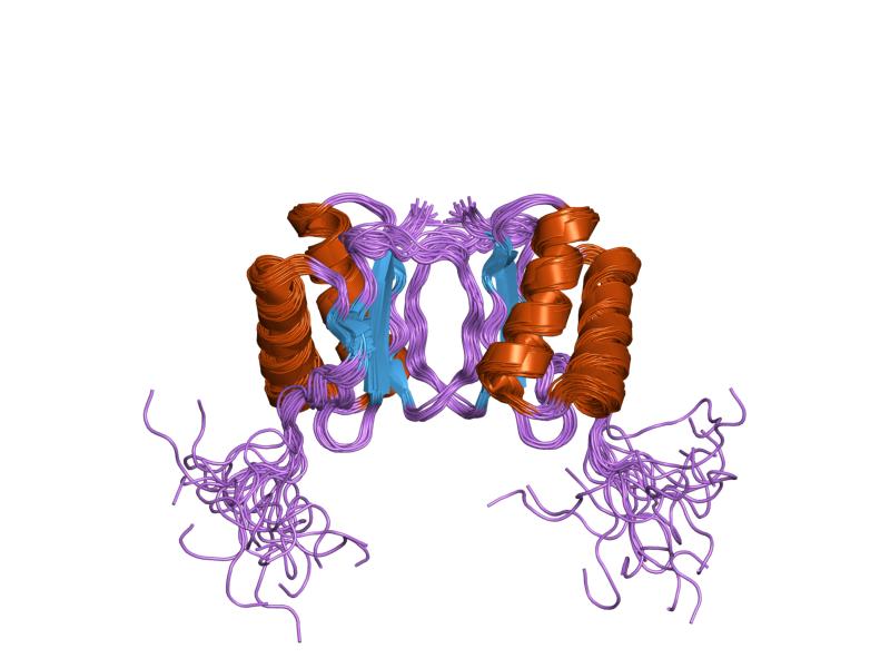
PDB gallery  1cmi: STRUCTURE OF THE HUMAN PIN/LC8 DIMER WITH A BOUND PEPTIDE
 1f3c: REFINED SOLUTION STRUCTURE OF 8KDA DYNEIN LIGHT CHAIN (DLC8)
 1f95: SOLUTION STRUCTURE OF DYNEIN LIGHT CHAIN 8 (DLC8) AND BIM PEPTIDE COMPLEX
 1f96: SOLUTION STRUCTURE OF DYNEIN LIGHT CHAIN 8 (DLC8) AND NNOS PEPTIDE COMPLEX
 1pwj: Structure of the Monomeric 8-kDa Dynein Light Chain and Mechanism of Domain Swapped Dimer Assembly
 1pwk: Structure of the Monomeric 8-kDa Dynein Light Chain and Mechanism of Domain Swapped Dimer Assembly
 1re6: Localisation of Dynein Light Chains 1 and 2 and their Pro-apoptotic Ligands
 1rhw: The solution structure of the pH-induced monomer of dynein light chain LC8 from Drosophila
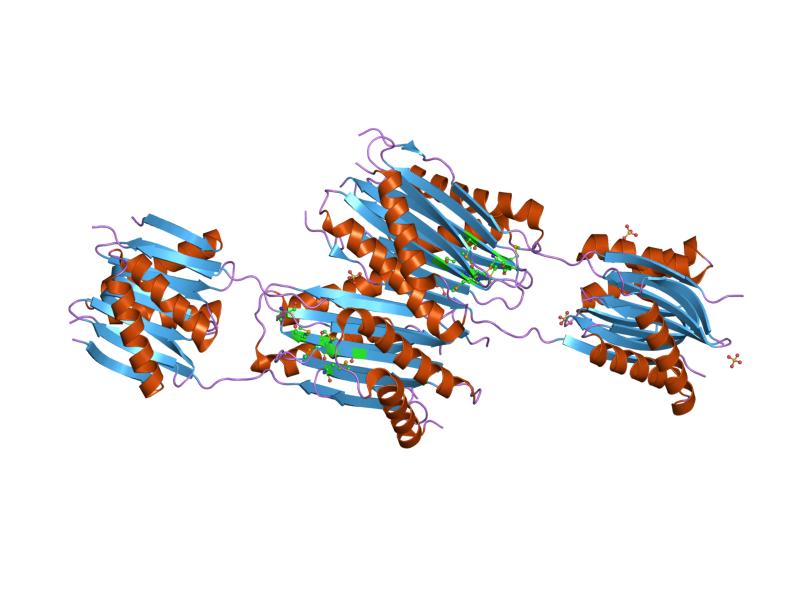
 2pg1: Structural analysis of a cytoplasmic dynein Light Chain-Intermediate Chain complexCategories:
- Human proteins
- Chromosome 12 gene stubs


Wikimedia Foundation. 2010.