- Main subunit of cytochrome c oxidase
-
Cytochrome C and Quinol oxidase polypeptide I 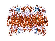
Structure of the 13-subunit oxidized cytochrome c oxidase.[1] Identifiers Symbol COX1 Pfam PF00115 InterPro IPR000883 PROSITE PDOC00074 SCOP 1occ TCDB 3.D.4 OPM family 4 OPM protein 1v55 Available protein structures: Pfam structures PDB RCSB PDB; PDBe PDBsum structure summary Cytochrome C and Quinol oxidase polypeptide I is main subunit of cytochrome c oxidase complex.
Cytochrome c oxidase (EC 1.9.3.1) is a key enzyme in aerobic metabolism. Proton pumping heme-copper oxidases represent the terminal, energy-transfer enzymes of respiratory chains in prokaryotes and eukaryotes. The CuB-heme a3 (or heme o) binuclear centre, associated with the largest subunit I of cytochrome c and ubiquinol oxidases (EC 1.10.3), is directly involved in the coupling between dioxygen reduction and proton pumping.[2][3] Some terminal oxidases generate a transmembrane proton gradient across the plasma membrane (prokaryotes) or the mitochondrial inner membrane (eukaryotes).
The enzyme complex consists of 3-4 subunits (prokaryotes) up to 13 polypeptides (mammals) of which only the catalytic subunit (equivalent to mammalian subunit I (CO I)) is found in all heme-copper respiratory oxidases. The presence of a bimetallic centre (formed by a high-spin heme and copper B) as well as a low-spin heme, both ligated to six conserved histidine residues near the outer side of four transmembrane spans within CO I is common to all family members.[4][5][6] In contrast to eukaryotes the respiratory chain of prokaryotes is branched to multiple terminal oxidases. The enzyme complexes vary in heme and copper composition, substrate type and substrate affinity. The different respiratory oxidases allow the cells to customize their respiratory systems according to a variety of environmental growth conditions.[2]
It has been shown that eubacterial quinol oxidase was derived from cytochrome c oxidase in Gram-positive bacteria and that archaebacterial quinol oxidase has an independent origin. A considerable amount of evidence suggests that proteobacteria (Purple bacteria) acquired quinol oxidase through a lateral gene transfer from Gram-positive bacteria.[2]
A related nitric oxide reductase (EC 1.7.99.7) exists in denitrifying species of archae and eubacteria and is a heterodimer of cytochromes b and c. Phenazine methosulphate can act as acceptor.
Subfamilies
- Cytochrome c oxidase cbb3-type, subunit I IPR004677
- Cytochrome o ubiquinol oxidase, subunit I IPR014207
- Cytochrome aa3 quinol oxidase, subunit I IPR014233
- Cytochrome c oxidase, subunit I bacterial type IPR014241
Examples
In humans, the main subunit of cytochrome c oxidase is encoded by the MT-CO1 gene.
References
- ^ Tsukihara T, Aoyama H, Yamashita E, et al. (May 1996). "The whole structure of the 13-subunit oxidized cytochrome c oxidase at 2.8 A". Science 272 (5265): 1136–44. Bibcode 1996Sci...272.1136T. doi:10.1126/science.272.5265.1136. PMID 8638158.
- ^ a b c Rumbley J, Gennis RB, Garcia-Horsman JA, Barquera B, Ma J (1994). "The superfamily of heme-copper respiratory oxidases". J. Bacteriol. 176 (18): 5587–5600. PMC 196760. PMID 8083153. http://www.pubmedcentral.nih.gov/articlerender.fcgi?tool=pmcentrez&artid=196760.
- ^ Glaser P, Villani G, Papa S, Capitanio N (1994). "The proton pump of heme-copper oxidases". Cell Biol. Int. 18 (5): 345–355. doi:10.1006/cbir.1994.1084. PMID 8049679.
- ^ Saraste M, Castresana J, Higgins DG, Lubben M (1994). "Evolution of cytochrome oxidase, an enzyme older than atmospheric oxygen". EMBO J. 13 (11): 2516–2525. PMC 395125. PMID 8013452. http://www.pubmedcentral.nih.gov/articlerender.fcgi?tool=pmcentrez&artid=395125.
- ^ Capaldi RA, Malatesta F, Darley-Usmar VM (1983). "Structure of cytochrome c oxidase". Biochim. Biophys. Acta 726 (2): 135–48. PMID 6307356.
- ^ Saraste M, Holm L, Wikstrom M (1987). "Structural models of the redox centres in cytochrome oxidase". EMBO J. 6 (9): 2819–2823. PMC 553708. PMID 2824194. http://www.pubmedcentral.nih.gov/articlerender.fcgi?tool=pmcentrez&artid=553708.
Categories:- Protein domains
- Protein families
- Transmembrane proteins

Wikimedia Foundation. 2010.