- Nerve injury
-
Nerve injury Classification and external resources 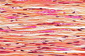
Micrograph of a nerve with a decrease in myelinated nerve fibres (pink) and an abnormal increase in fibrous tissue (yellow), as may be seen in nerve injuries. HPS stain.ICD-10 T14.4 Nerve injury is injury to nervous tissue. There is no single classification system that can describe all the many variations of nerve injury. Most systems attempt to correlate the degree of injury with symptoms, pathology and prognosis.[citation needed] In 1941, Seddon introduced a classification of nerve injuries based on three main types of nerve fiber injury and whether there is continuity of the nerve.[1]
Contents
Types
Neurapraxia
This is the least severe form of nerve injury, with complete recovery. In this case, the actual structure of the nerve remains intact, but there is an interruption in conduction of the impulse down the nerve fiber. Most commonly, this involves compression of the nerve or disruption to the blood supply (ischemia). There is a temporary loss of function which is reversible within hours to months of the injury (the average is 6–9 weeks). Wallerian degeneration does not occur, so recovery does not involve actual regeneration. There is frequently greater involvement of motor than sensory function with autonomic function being retained. In electrodiagnostic testing with nerve conduction studies, there is a normal compound motor action potential amplitude distal to the lesion at day 10, and this indicates a diagnosis of mild neuropraxia instead of axonotmesis or neurotmesis.[2]
Axonotmesis
This is a more severe nerve injury with disruption of the neuronal axon, but with maintenance of the myelin sheath.[clarification needed] This type of nerve damage may cause paralysis of the motor, sensory, and autonomic. Mainly seen in crush injury.
If the force creating the nerve damage is removed in a timely fashion, the axon may regenerate, leading to recovery. Electrically, the nerve shows rapid and complete degeneration, with loss of voluntary motor units. Regeneration of the motor end plates will occur, as long as the endoneural tubules are intact.
Axonotmesis involves loss of the relative continuity of the axon and its covering of myelin,[clarification needed] but preservation of the connective tissue framework of the nerve ( the encapsulating tissue, the epineurium and perineurium, are preserved ). Because axonal continuity is lost, Wallerian degeneration occurs. Electromyography ( EMG ) performed 2 to 4 weeks later shows fibrillations and denervation potentials in musculature distal to the injury site. Loss in both motor and sensory spines is more complete with axonotmesis than with neurapraxia, and recovery occurs only through regenerations of the axons, a process requiring time.
Axonotmesis is usually the result of a more severe crush or contusion than neurapraxia, but can also occur when the nerve is stretched (without damage to the epineurium). There is usually an element of retrograde proximal degeneration of the axon, and for regeneration to occur, this loss must first be overcome. The regeneration fibers must cross the injury site and regeneration through the proximal or retrograde area of degeneration may require several weeks. Then the neuritis tip progresses down the distal site, such as the wrist or hand. Proximal lesion may grow distally as fast as 2 to 3 mm per day and distal lesion as slowly as 1.5 mm per day. Regeneration occurs over weeks to years.
Neurotmesis
Neurotmesis is the most severe lesion with potential of recovering. It occurs on severe contusion, stretch, laceration, or Local Anesthetic Toxicity. Not only the axon, but the encapsulating connective tissue lose their continuity. The last (extreme) degree of neurotmesis is transsection, but most neurotmetic injuries do not produce gross loss of continuity of the nerve but rather internal disruption of the architecture of the nerve sufficient to involve perineurium and endoneuruim as well as axons and their covering. Denervation changes recorded by EMG are the same as those seen with axonotmetic injury. There is a complete loss of motor, sensory and autonomic function. If the nerve has been completely divided, axonal regeneration causes a neuroma to form in the proximal stump. For neurotmesis, it is better to use a new more complete classification called the Sunderland System.
Regeneration
Main article: NeuroregenerationPhysiological mechanisms or neuroregeneration may include remyelination, generation of new neurons, glia, axons, myelin or synapses. Neuroregeneration differs between the Peripheral Nervous System (PNS) and the Central Nervous System (CNS) by the functional mechanisms and especially, the extent and speed.
Surgery can be done in case a peripheral nerve has become cut or otherwise divided. Recovery of a nerve after surgical repair depends mainly on the age of the patient. Young children can recover close-to-normal nerve function.[3] In contrast, a patient over 60 years old with a cut nerve in the hand would expect to recover only protective sensation, that is, the ability to distinguish hot/cold or sharp/dull.[3] Many other factors also affect nerve recovery.[3]
In contrast, repair after damage to the central nervous system is limited.
See also
- Brain injury (disambiguation)
- Neuroregeneration
- Peripheral nerve
- Peripheral nerve injury
- Seddon's classification
- Wallerian degeneration
References
- ^ Seddon, H.J.: Classification of nerve injuries, British Medical Journal, 2:237, 1942.
- ^ Faubel, C: Nerve Injury Classifications – Seddon’s and Sunderland’s. www.ThePainSource.com, 2010.
- ^ a b c The Southern Orthopaedic Association > Patient Education: Nerve Repair and Grafting in the Upper Extremity 2006. Retrieved on Jan 12, 2009
Neurotrauma (S06, Sx4, T09.3–4, 850–854, 950–957) Traumatic brain injury Intracranial hemorrhage/hematoma: intra-axial (Intraparenchymal hemorrhage, Intraventricular hemorrhage) · extra-axial (Subdural hematoma, Epidural hematoma, Subarachnoid hemorrhage)Spinal cord injury PNS Nerve injury (Peripheral nerve injury), Wallerian degenerationCategories:- Peripheral nervous system disorders
- Neurotrauma

Wikimedia Foundation. 2010.