- Myiasis
-
Myiasis Classification and external resources 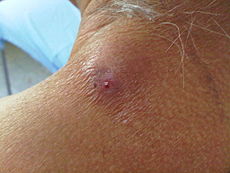
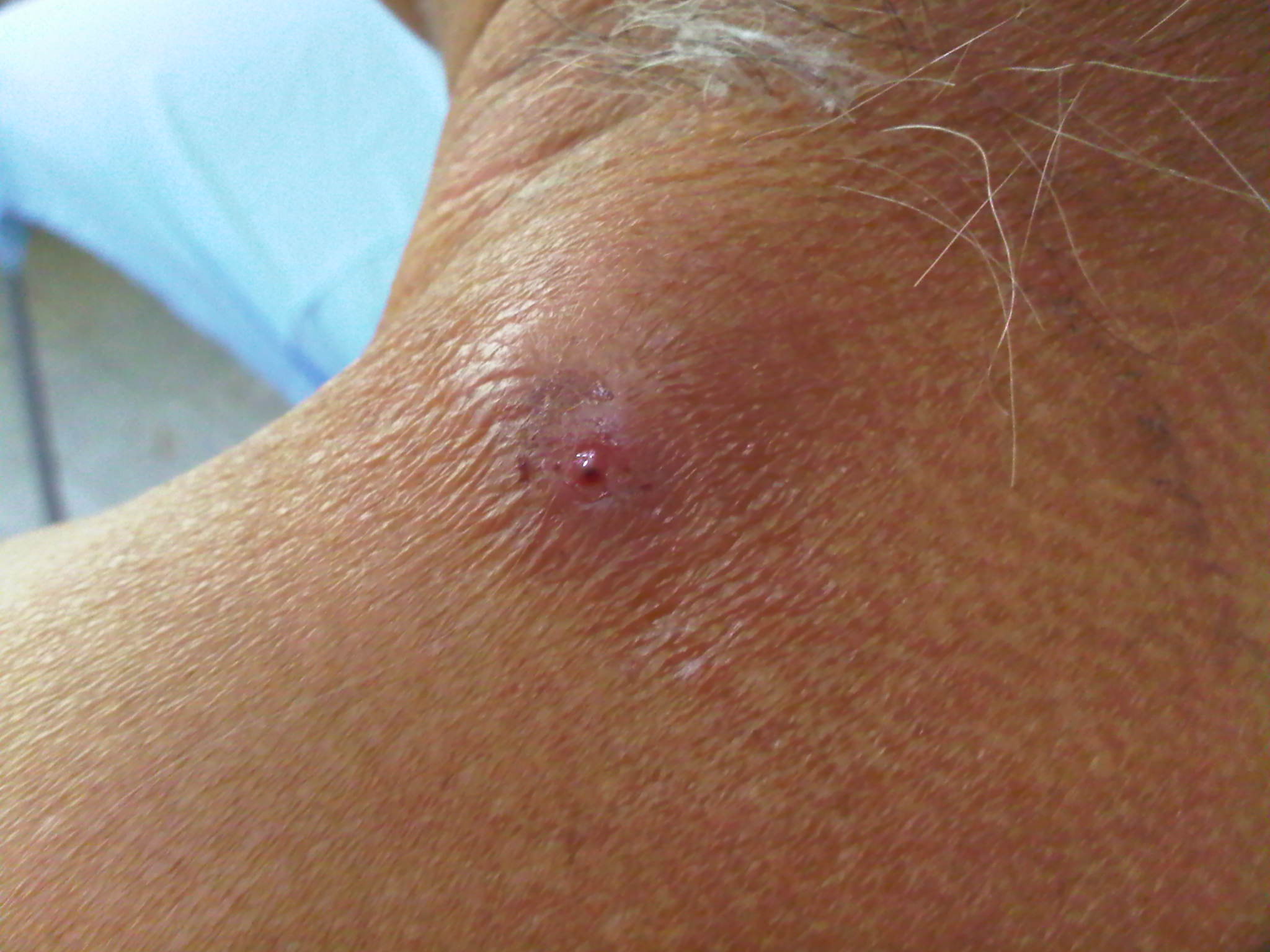
Cutaneous myiasis in the neck of a humanICD-10 B87 ICD-9 134.0 DiseasesDB 29588 MeSH D009198 Myiasis (
 /ˈmaɪ.əsɨs/ or /maɪˈaɪ.əsɨs/) is a general term for infection by parasitic fly larvae feeding on the host's necrotic or living tissue. Colloquialisms for myiasis include flystrike, blowfly strike, and fly-blown. In Greek, "myia" means fly.
Myiasis is a serious problem for livestock industries, causing severe economic losses worldwide.[1] Although infestation by fly larvae is much more prevalent in animals, it is a relatively frequent occurrence in humans in rural, tropical and subtropical regions.[2]
Myiasis can come in all sorts of variations, depending on the fly species and where the larvae are located. Some flies may lay eggs in open wounds, other larvae may invade unbroken skin or enter the body through the nose or ears, and still others may be swallowed if the eggs are deposited on the lips or on food.[2]
Contents
History of discovery
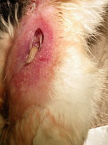 Myiasis in a cat's flesh
The Reverend Frederick William Hope coined the term myiasis in 1840 to refer to diseases resulting from dipterous larvae as opposed to those caused by other insect larvae (the term for this was scholechiasis). Hope described several cases of myiasis from Jamaica caused by unknown larvae, one of which resulted in death.[3]
Even though the term myiasis was first used in 1840, the disease has been around for far longer than that. Ambroise Paré, the chief surgeon to Charles IX and Henri III, observed that maggots often infested open wounds.[4]
Life cycle
Myiasis, known as blowfly strike, is a common disease in sheep, especially in areas where there are hot and wet conditions. The life cycle in sheep is typical of the disease. The female flies lay their eggs on the sheep in damp, protected areas of the body that are soiled with urine and faeces, mainly the sheep's breech (buttocks). It takes approximately eight hours to a day for the eggs to hatch, depending on the conditions. This results in sores as the larvae lacerate the skin; this is the primary reason for the early removal of lambs' tails. The larvae then tunnel into the host's tissue, causing irritating lesions. After about the second day, bacterial infection occurs and, if left untreated, causes toxemia or septicemia. This leads to anorexia and weakness and, if untreated, will lead to death. Blowfly strike accounts for over A$170 million a year in losses in the Australian sheep industry so preventive measures such as mulesing are practised. Infestation of vulvar area with larvae and maggots is called vulvar myiasis.
Classifications
German entomologist Fritz Zumpt describes myiasis as "the infestation of live human and vertebrate animals with dipterous larvae, which at least for a period, feed on the host's dead or living tissue, liquid body substances, or ingested food". For modern purposes however, this is too vague. For example, feeding on dead tissue is not generally a problem except when larvae such as those of flies in the familiy Piophilidae attack stored food such as cheese or preserved meats; such activity suggests saprophagy rather than parasitism; it even may be medically beneficial in maggot debridement therapy (MDT).
Currently myiasis commonly is classified according to aspects relevant to the case in question:
- The classical description of myiasis is according to the part of the host that is infected. This is the classification used by ICD-10. For example:[5]
- dermal
- sub-dermal
- cutaneous (B87.0)
- Creeping, where larvae burrow through or under the skin
- Furuncular, where a larva remains in one spot, causing a boil-like lesion
- nasopharyngeal nose, sinuses or pharynx. (B87.3)
- Ophthalmic or ocular in or about the eye (B87.2)
- Auricular in or about the ear
- gastric, rectal, or intestinal/enteric for the appropriate part of the digestive system(B87.8)
- urogenital (B87.8).
- Another aspect is the relationship between the host and the parasite and provides insight into the biology of the fly species causing the myiasis and its likely effect. Thus the myiasis is described as either:[5]
- Obligatory, where the parasite cannot complete its life cycle without its parasitic phase, which may be Specific, Semispecific, or Opportunistic.
- Facultative, incidental, or accidental, where it is not essential to the life cycle of the parasite; perhaps a normally free-living larva accidentally gained entrance to the host.[2]
Accidental myiasis commonly is enteric, resulting from swallowing eggs or larvae with one's food. The effect is called pseudomyiasis.[6] One traditional cause of pseudomyiasis was the eating of maggots of cheese flies in cheeses such as Stilton. Depending on the species present in the gut, pseudomyiasis may cause significant medical symptoms, but it is likely that most cases pass unnoticed.
Vectors in humans
There are three main fly families causing economically important myiasis in livestock and also, occasionally, in humans:
- Calliphoridae (blowflies)
- Oestroidea (botflies)
- Sarcophagidae (fleshflies)
Other families occasionally involved are:
- Anisopodidae
- Piophilidae
- Stratiomyidae
- Syrphidae
Specific myiasis
Caused by flies that need a host for larval development
- Dermatobia hominis (human botfly)
- Cordylobia anthropophaga (tumbu fly)
- Oestrus ovis (sheep botfly)
- Hypoderma spp. (cattle botflies or ox warbles)
- Gasterophilus spp. (horse botfly)
- Cochliomyia hominivorax (new world screwworm fly)
- Chrysomya bezziana (old world screwworm fly)
- Auchmeromyia senegalensis (Congo floor maggot)
- Cuterebra spp. (rodent and rabbit botfly)
Semispecific myiasis
Caused by flies that usually lay their eggs in decaying animal or vegetable matter, but that can develop in a host if open wounds or sores are present
- Lucilia spp. (green-bottle fly)
- Cochliomyia spp. (blue-bottle fly)
- Phormia spp. (black-bottle fly)
- Calliphora spp. (blowfly)
- Sarcophaga spp. (flesh fly or sarcophagids)
Flesh flies, or sarcophagids, can cause intestinal myiasis in humans if the females lay their eggs on meat or fruit.
Accidental myiasis
Also called pseudomyiasis. Caused by flies that have no preference or need to develop in a host but that will do so on rare occasions. Transmission occurs through accidental deposit of eggs on oral or genitourinary openings, or by swallowing eggs or larvae that are on food.
- Musca domestica (housefly)
- Fannia spp. (latrine flies)
- Eristalis tenax (rat-tailed maggots)
- Muscina spp.
The adult flies are not parasitic, but when they lay their eggs in open wounds and these hatch into their larval stage (also known as maggots or grubs), the larvae feed on live and/or necrotic tissue, causing myiasis to develop. They may also be ingested or enter through other body apertures.
Clinical presentation in humans
How myiasis affects the human body depends on where the larvae are located. Larvae may infect necrotic (dead) or living tissue in various sites: the skin, eyes, ears, stomach and intestinal tract, or in genitourinary sites.[7] They may invade open wounds and lesions or unbroken skin. Some enter the body through the nose or ears. Larvae or eggs can reach the stomach or intestines if they are swallowed with food and cause gastric or intestinal myiasis.[2]
Several different presentations of myiasis and their symptoms:[2]
Syndrome Symptoms Cutaneous Myiasis Painful, slow-developing ulcers or furuncle- (boil-) like sores that can last for a prolonged period. Nasal Myiasis Obstruction of nasal passages and severe irritation. In some cases facial edema and fever can develop. Death is not uncommon. Aural Myiasis Crawling sensations and buzzing noises. Smelly discharge is sometimes present. If located in the middle ear, larvae may get to the brain. Ophthalmomyiasis Fairly common, this causes severe irritation, edema, and pain. Nosocomial Myiasis refers to myiasis in a hospital setting. It is quite frequent, as patients with open wounds or sores can be infested if flies are present. To prevent nosocomial myiasis, hospital rooms must be kept free of flies.
Human ophthalmomyiasis, both external and internal, has been caused by botfly larvae.[8]
Diagnostics
Myiasis is often misdiagnosed in the United States because it is extremely rare and its symptoms are not specific. Intestinal myiasis and urinary myiasis are especially difficult to diagnose.[2]
Clues that myiasis may be present include recent travel to an endemic area, one or more non-healing lesions on the skin, itchiness, movement under the skin or pain, discharge from a central punctum (tiny hole), or a small, white structure protruding from the lesion.[9] Serologic testing has also been used to diagnose the presence of botfly larvae in human ophthalmomyiasis.[8]
Control methods
The first control method is preventive and aims to eradicate the adult flies before they can cause any damage and is called vector control. The second control method is the treatment once the infestation is present, and concerns the infected animals (or humans).
Prevention
The principal control method of adult populations of myiasis inducing flies involves insecticide applications in the environment where the target livestock is kept. Organophosphorus or organochlorine compounds may be used, usually in a spraying formulation. One alternative prevention method is the SIT (Sterile Insect Technique) where a significant number of artificially reared sterilized (usually through irradiation) male flies are introduced. The male flies compete with wild bred males for females in order to copulate and thus cause females to lay batches of unfertilized eggs which can't develop into the larval stage.
One prevention method involves removing the environment most favourable to the flies, such as by docking (removal of the tail). Another example is the crutching of sheep, which involves the removal of wool from around the tail and between the rear legs, which is a favourable environment for the larvae. Another more permanent practice which is used in some countries is mulesing, where skin is removed from young animals to tighten remaining skin – leaving it less prone to fly attack.[10] PETA have been campaigning to have farmers cease mulesing. Celebrities such as Pink, Toni Collette and Chrissie Hynde have also participated in PETA's campaign against the mulesing practice;[11] However, Collette has since changed her stance[12] and Pink has said she was misled by PETA and had not done enough research herself into mulesing.
To prevent myiasis in humans, there is a need for general improvement of sanitation, personal hygiene, and extermination of the flies by insecticides. Clothes should be washed thoroughly, dried away from flies, and ironed. The heat of the iron kills the eggs of myiasis-causing flies.[9]
Treatment
This applies once an infestation is underway. First the larvae must be eliminated through pressure around the lesion and the use of forceps. Secondly the wound must be cleaned and disinfected. Further control is necessary to avoid further reinfestation.
It is also possible to treat livestock with the use of slow release boluses containing ivermectin which can provide long term protection against the larvae development.
Sheep may be dipped, which involves drenching the sheep in insecticide to prevent the growth of the larvae.
Use of myiasitic maggots in medicine
Main article: Maggot therapyThroughout time maggots have been used as medicine in order to clean out necrotic wounds, a method known as maggot therapy.
Fly larvae that feed on dead tissue can clean wounds and may reduce bacterial activity and the chance of a secondary infection. They dissolve dead tissue by secreting digestive enzymes onto the wound as well as actively eat the dead tissue with “mouth hooks,” two probing appendages near their toothless mouth.[13] Maggot therapy – also known as maggot debridement therapy (MDT), larval therapy, larva therapy, or larvae therapy – is the intentional introduction by a health care practitioner of live, disinfected green bottle fly maggots or larvae into the non-healing skin and soft tissue wound(s) of a human or other animal for the purpose of selectively cleaning out only the necrotic (dead) tissue within a wound in order to promote wound healing.
Maggot therapy has a long history. The indigenous people of Australia used maggot therapy, as well as the Hill Peoples of Northern Burma, and possibly the Mayans of Central America.[2] Surgeons in Napoleon’s armies recognized that wounded soldiers with myiasis were more likely to survive than those without the infestation. In the American Civil War, army surgeons treated wounds by allowing blowfly maggots to clean away the decayed tissue.
Dr. William Baer, an orthopedic surgeon at Johns Hopkins during the late 1920s, used maggot therapy to treat a series of patients with osteomyelitis, an infection of bone or bone marrow and published his findings. Dr. Baer first got the idea when, during World War I, two soldiers presented to him with broken femurs after having lain on the ground for seven days without food and water. Dr. Baer could not figure out why neither man had a fever or signs of sepsis, and he got an unexpected surprise: “On removing the clothing from the wounded part, much was my surprise to see the wound filled with thousands and thousands of maggots, apparently those of the blow fly. The sight was very disgusting and measures were taken hurriedly to wash out these abominable looking creatures.” However, he then realized that the wounds were filled with “beautiful pink granulation tissue” and were healing well. Dr. Baer was onto something that many had discovered before him: the healing power of maggots.[14]
Maggot therapy was common in the United States during the 1930s. However, during the second half of the twentieth century, after the introduction of antibiotics, maggot therapy was only used as a last resort for very serious wounds.[2] Lately maggots have been making a comeback due to the increased resistance of bacteria to antibiotics.
Although maggot therapy has been used in the US for the past 80 years, it was approved by the FDA as a “medical device” only in 2004 (along with leeches in the same year).[15] Maggots were the first live organism to be marketed in the US according to FDA regulations, and are approved for treating neuropathic (diabetic) foot ulcers, pressure ulcers, venous stasis ulcers, and traumatic and post-surgical wounds that are unresponsive to conventional therapies. Before this, maggots were being used but were unregulated. Richard Sherman, a doctor in Irvine, CA, is the most well-known maggot therapy advocate and began treating patients with maggots in 1990.[16] He is now the laboratory director of Monarch Labs, the first and only commercial producer of medical grade maggots. This lab has provided maggots and maggot therapy supplies since 1935.[17]
Today, there is an ever-increasing demand for medical maggots. In the US, demand for these fly larvae has increased by 20% over the last ten years, and doubled since the FDA ruling. Maggot therapy is now used in more than 300 sites across the country.[13] The American Medical Association and Centers for Medicare and Medicaid Services recently clarified the reimbursement guidelines to the wound care community for medicinal maggots, and this therapy may soon be covered by insurance.[18] The larvae of the green bottle fly (a type of blow-fly) are now used exclusively for this purpose, since they preferentially devour only necrotic tissue, leaving healthy tissue intact. This is an important distinction, as most other major varieties of myiasitic fly larvae attack both live and dead wound tissue indiscriminately, effectively negating their benefit in non-harmful wound debridement. Medicinal maggots are placed on the wound and covered with a sterile dressing of gauze and nylon mesh. Too many larvae placed on the wound could result in healthy tissue being eaten.[4]
See also
References
- ^ Otranto, Domenico. “The immunology of myiasis: parasite survival and host defense strategies.” Trends in Parasitology 17 (2001): 4.
- ^ a b c d e f g h John, David and Petri, William. Markell and Voge’s Medical Parasitology: 9th Edition. Missouri: Saunders Elsevier, 2006. p. 328-334.
- ^ “Introduction to Myiasis.” The Natural History Museum of London. <http://www.nhm.ac.uk/research-curation/research/projects/myiasis-larvae/intro-myiasis/index.html>.
- ^ a b Sherman, RA, Hall, MJR, and Thomas, S. “Medicinal Maggots: An ancient remedy for some contemporary afflictions.” Annual Review of Entomology 45 (2000): 55-81.
- ^ a b Janovy, John; Schmidt, Gerald D.; Roberts, Larry S. (1996). Gerald D. Schmidt & Larry S. Roberts' Foundations of parasitology. Dubuque, Iowa: Wm. C. Brown. ISBN 0-697-26071-2.
- ^ Zumpt, Fritz Konrad Ernst; Myiasis in man and animals in the old world. Butterworth 1965
- ^ Ockenhouse, Christian, Samlaska, Curt, Benson, Paul, Roberts, Lyman, Eliasson, Arn, Malane, Susan, and Menich, Mark. “Cutaneous myiasis caused by the African tumbu fly.” Archives of Dermatology 126 (1990):199-202.
- ^ a b Lagacé-Wiens, Philippe (2008). "Human Ophthalmomyiasis Interna Caused by Hypoderma tarandi, Northern Canada". Emerging Infectious Diseases 14 (1). http://www.cdc.gov/eid/content/14/1/64.htm. Retrieved 22 June 2011.
- ^ a b Adisa, Charles and Mbanaso, Augustus. “Furuncular myiasis of the breast caused by the larvae of the Tumbu fly (Cordylobia anthropophaga).” BMC Surgery 4 (2004): 5.
- ^ "Standard Operating Procedures - sheep Mulesing". teacher's notes. New South Wales Department of Primary Industries. March 8, 2004. http://www.agric.nsw.gov.au/reader/aw-sheep/13-mulesing.htm. Retrieved 2007-01-09.
- ^ "Pink angers Australian government". BBC News. 20 December 2006. http://news.bbc.co.uk/2/hi/entertainment/6196691.stm. Retrieved 2007-01-09.
- ^ Actress backs down on call for mulesing ban - 18/07/2005
- ^ a b Greer, Kathleen. “Age-old therapy gets new approval.” Advances in Skin & Wound Care. January/February 2005.
- ^ Baer, William. “The treatment of chronic osteomyelitis with the maggot (larva of the blow fly).” Journal of Bone and Joint Surgery 13 (1931): 438-475.
- ^ Rubin, Rita. “Maggots and leeches: Good medicine.” USA Today 7 July 2004 < http://www.usatoday.com/news/health/2004-07-07-leeches-maggots_x.htm>.
- ^ Rubin, Rita. “Maggots and leeches: Good medicine.” USA Today 7 July 2004 < http://www.usatoday.com/news/health/2004-07-07-leeches-maggots_x.htm>.
- ^ Maggot Therapy Project. Ed. Ronald Sherman. 2009. University of California Irvine Department of Pathology. 2 Feb. 2009 <http://www.healthaffairs.uci.edu/som/pathology/sherman/home_pg.htm>.
- ^ Lloyd, Robin. “ Insurance may soon cover maggot therapy.” MSNBC 19 Nov. 2008 <http://www.msnbc.msn.com/id/27808424/>.
External links
- Myiasis reviewed and published by WikiVet
- University of Sydney
- fly blown sheep
- Human oral myiasis, two cases and general remarks
- pdf A collective analysis of 54 cases of human myiasis in China from 1995–2001.Chinese Medical Journal
- NHM Identification key to myiasis causing fly larvae.
Infectious diseases – Parasitic disease: ectoparasitic infestation / arthropod (B85–B89, 132–134) Insecta Pediculus humanus corporis/Pediculus humanus capitis (Pediculosis, Head-louse infestation, Pediculosis corporis)Cimex (Cimicosis)Dermatobia hominis/Cordylobia anthropophaga (Myiasis)Tunga penetrans (Tungiasis)Arachnida Trombidiformes: Trombicula (Trombiculosis, Chigger bite) · Demodex brevis/Demodex folliculorum (Demodicosis, Demodex mite bite) · Pyemotes herfsi · Cheyletiella (Cheyletiellosis)Sarcoptiformes: Sarcoptes scabiei (Scabies)Dermanyssus gallinae · Liponyssoides sanguineusTick infestationCrustacea Linguatula serrata (Linguatulosis) · Porocephalus crotali/Armillifer armillatus (Porocephaliasis)M: IFT
helm,arth (acar)
helm, arth (lice), zoon
helm, arth
Categories:- Sheep and goat diseases
- Arthropod infestations
- Veterinary entomology
- Parasitic infestations, stings, and bites of the skin
- The classical description of myiasis is according to the part of the host that is infected. This is the classification used by ICD-10. For example:[5]

Wikimedia Foundation. 2010.