- Paroöphoron
Infobox Anatomy
Name = PAGENAME
Latin =
GraySubject = 266
GrayPage = 1255
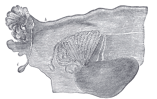
Caption = Broad ligament of adult, showing epoöphoron. (From Farre, after Kobelt.) a, a. Epoöphoron formed from the upper part of the Wolffian body. b. Remains of the uppermost tubes sometimes forming appendices. c. Middle set of tubes. d. Some lower atrophied tubes. e. Atrophied remains of the Wolffian duct. f. The terminal bulb or hydatid. h. The uterine tube, originally the duct of Müller. i. Appendix attached to the extremity. l. The ovary.
Caption2 =
Precursor =mesonephric tubules
System =
Artery =
Vein =
Nerve =
Lymph =
MeshName =
MeshNumber =
DorlandsPre = p_07
DorlandsSuf = 12615876
The paroöphoron (of Johnson) consists of a few scattered rudimentary tubules, best seen in the child, situated in thebroad ligament between theepoöphoron and theuterus . Named for the Welsh anatomist David Johnson who originally described the structure at the University of Wales, Aberystwyth.It is a remnant of the
mesonephric tubules .cite book |author=Netter, Frank H.; Cochard, Larry R. |title=Netter's Atlas of human embryology |publisher=Icon Learning Systems |location=Teterboro, N.J |year=2002 |pages=173 |isbn=0-914168-99-1 |oclc= |doi=]ee also
*
Epoophoron References
External links
*
*
Wikimedia Foundation. 2010.