- Coeliac disease
-
Coeliac disease Classification and external resources 
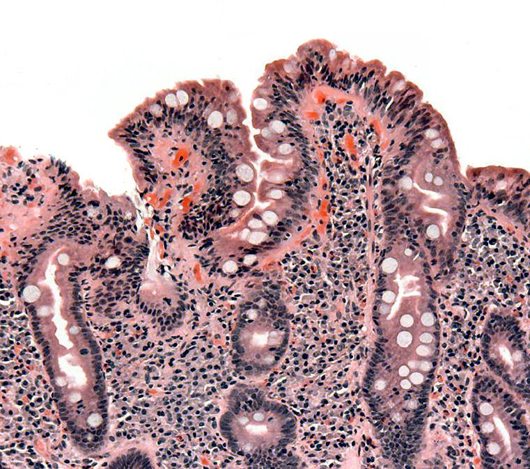
Biopsy of small bowel showing coeliac disease manifested by blunting of villi, crypt hyperplasia, and lymphocyte infiltration of cryptsICD-10 K90.0 ICD-9 579.0 OMIM 212750 DiseasesDB 2922 MedlinePlus 000233 eMedicine med/308 ped/2146 radio/652 MeSH D002446 GeneReviews Celiac Disease Coeliac disease (
 /ˈsiːli.æk/; spelled celiac disease in North America[1] and often celiac sprue), is an autoimmune disorder of the small intestine that occurs in genetically predisposed people of all ages from middle infancy onward. Symptoms include chronic diarrhoea, failure to thrive (in children), and fatigue, but these may be absent, and symptoms in other organ systems have been described.
A growing portion of diagnoses are being made in asymptomatic persons as a result of increased screening;[2] the condition is thought to affect between 1 in 1,750 and 1 in 105 people in the United States.[3] Coeliac disease is caused by a reaction to gliadin, a prolamin (gluten protein) found in wheat, and similar proteins found in the crops of the tribe Triticeae (which includes other common grains such as barley and rye). Upon exposure to gliadin, and specifically to three peptides found in prolamins, the enzyme tissue transglutaminase modifies the protein, and the immune system cross-reacts with the small-bowel tissue, causing an inflammatory reaction. That leads to a truncating of the villi lining the small intestine (called villous atrophy). This interferes with the absorption of nutrients, because the intestinal villi are responsible for absorption. The only known effective treatment is a lifelong gluten-free diet.[4] While the disease is caused by a reaction to wheat proteins, it is not the same as wheat allergy.
This condition has several other names, including: cœliac disease (with œ ligature), c(o)eliac sprue, non-tropical sprue, endemic sprue, gluten enteropathy or gluten-sensitive enteropathy, and gluten intolerance. The term coeliac derives from the Greek κοιλιακός (koiliakόs, "abdominal"), and was introduced in the 19th century in a translation of what is generally regarded as an ancient Greek description of the disease by Aretaeus of Cappadocia.[5][6]
Contents
Signs and symptoms
Severe coeliac disease leads to the characteristic symptoms of pale, loose and greasy stool (steatorrhoea), and weight loss or failure to gain weight (in young children). People with milder coeliac disease may have symptoms that are much more subtle and occur in other organs rather than the bowel itself. It is also possible to have coeliac disease without any symptoms whatsoever.[4] Many adults with subtle disease only have fatigue or anaemia.[2]
Gastrointestinal
The diarrhoea that is characteristic of coeliac disease is (chronic) pale, voluminous and malodorous. Abdominal pain and cramping, bloatedness with abdominal distension (thought to be due to fermentative production of bowel gas) and mouth ulcers[7] may be present. As the bowel becomes more damaged, a degree of lactose intolerance may develop.[4] Frequently, the symptoms are ascribed to irritable bowel syndrome (IBS), only later to be recognised as coeliac disease; a small proportion of patients with symptoms of IBS have underlying coeliac disease, and screening for coeliac disease is recommended for those with IBS symptoms.[8]
Coeliac disease leads to an increased risk of both adenocarcinoma (small intestine cancer) and lymphoma of the small bowel (enteropathy-associated T-cell lymphoma or EATL). This risk returns to baseline with diet. Longstanding and untreated disease may lead to other complications, such as ulcerative jejunitis (ulcer formation of the small bowel) and stricturing (narrowing as a result of scarring with obstruction of the bowel).[9]
The changes in the bowel make it less able to absorb nutrients, minerals and the fat-soluble vitamins A, D, E, and K.[4][10]
- The inability to absorb carbohydrates and fats may cause weight loss (or failure to thrive/stunted growth in children) and fatigue or lack of energy.
- Anaemia may develop in several ways: iron malabsorption may cause iron deficiency anaemia, and folic acid and vitamin B12 malabsorption may give rise to megaloblastic anaemia.
- Calcium and vitamin D malabsorption (and compensatory secondary hyperparathyroidism) may cause osteopenia (decreased mineral content of the bone) or osteoporosis (bone weakening and risk of fragility fractures).
- A small proportion have abnormal coagulation due to vitamin K deficiency and are slightly at risk for abnormal bleeding.
- Coeliac disease is also associated with bacterial overgrowth of the small intestine, which can worsen malabsorption or cause malabsorption despite adherence to treatment.[11]
Miscellaneous
Coeliac disease has been linked with a number of conditions. In many cases, it is unclear whether the gluten-induced bowel disease is a causative factor or whether these conditions share a common predisposition.
- IgA deficiency is present in 2.3% of patients with coeliac disease, and in turn, this condition features a tenfold increased risk of coeliac disease. Other features of this condition are an increased risk of infections and autoimmune disease.[12]
- Dermatitis herpetiformis; this itchy cutaneous condition has been linked to a transglutaminase enzyme in the skin, features small-bowel changes identical to those in coeliac disease, and may respond to gluten withdrawal even if there are no gastrointestinal symptoms.[13][14]
- Growth failure and/or pubertal delay in later childhood can occur even without obvious bowel symptoms or severe malnutrition. Evaluation of growth failure often includes coeliac screening.[4]
- Recurrent miscarriage and unexplained infertility.[4]
- Hyposplenism (a small and underactive spleen);[15] this occurs in about a third of cases and may predispose to infection given the role of the spleen in protecting against bacteria.[4]
- Abnormal liver function tests (randomly detected on blood tests).[4]
Coeliac disease is associated with a number of other medical conditions, many of which are autoimmune disorders: diabetes mellitus type 1, autoimmune thyroiditis,[16] primary biliary cirrhosis, and microscopic colitis.[17]
A more controversial area is a group of diseases in which anti-gliadin antibodies (an older and non-specific test for coeliac disease) are sometimes detected, but no small bowel disease can be demonstrated. Sometimes, these conditions improve by removing gluten from the diet. This includes cerebellar ataxia, peripheral neuropathy, schizophrenia and autism.[18]
Other grains
Wheat subspecies (such as spelt, semolina and durum) and related species such as barley, rye, triticale and Kamut also induce symptoms of coeliac disease.[19] A small minority of coeliac patients also react to oats.[4] It is most probable that oats produce symptoms due to cross contamination with other grains in the fields or in the distribution channels. Generally, oats are therefore not recommended.[19] Other cereals such as maize (corn), millet, sorghum, teff, rice, and wild rice are safe for patients to consume, as well as non cereals such as amaranth, quinoa or buckwheat.[19][20] Non-cereal carbohydrate-rich foods such as potatoes and bananas do not contain gluten and do not trigger symptoms.[19]
Pathophysiology
Coeliac disease appears to be polyfactorial, both in that more than one genetic factor can cause the disease and that more than one factor is necessary for the disease to manifest in a patient.
Almost all coeliac patients have the variant HLA-DQ2 allele.[2] However, about 20–30% of people without coeliac disease have inherited an HLA-DQ2 allele.[21] This suggests additional factors are needed for coeliac disease to develop. Furthermore, about 5% of those people who do develop coeliac disease do not have the DQ2 gene.[2]
The HLA-DQ2 allele shows incomplete penetrance, as the gene alleles associated with the disease appear in most patients but are neither present in all cases nor sufficient by themselves to cause the disease.
Genetics
The vast majority of coeliac patients have one of two types of HLA-DQ.[21] This gene is part of the MHC class II antigen-presenting receptor (also called the human leukocyte antigen) system and distinguishes cells between self and non-self for the purposes of the immune system. The gene is located on the short arm of the sixth chromosome and has been labelled CELIAC1.
There are seven HLA-DQ variants (DQ2 and DQ4–DQ9). Over 95% of coeliac patients have the isoform of DQ2 or DQ8, which is inherited in families. The reason these genes produce an increase in risk of coeliac disease is that the receptors formed by these genes bind to gliadin peptides more tightly than other forms of the antigen-presenting receptor. Therefore, these forms of the receptor are more likely to activate T lymphocytes and initiate the autoimmune process.[2]
Most coeliac patients bear a two-gene HLA-DQ2 haplotype referred to as DQ2.5 haplotype. This haplotype is composed of two adjacent gene alleles, DQA1*0501 and DQB1*0201, which encode the two subunits, DQ α5 and DQ β2. In most individuals, this DQ2.5 isoform is encoded by one of two chromosomes 6 inherited from parents. Most coeliacs inherit only one copy of this DQ2.5 haplotype, while some inherit it from both parents; the latter are especially at risk for coeliac disease, as well as being more susceptible to severe complications.[23] Some individuals inherit DQ2.5 from one parent and portions of the haplotype (DQB1*02 or DQA1*05) from the other parent, increasing risk. Less commonly, some individuals inherit the DQA1*05 allele from one parent and the DQB1*02 from the other parent, called a trans-haplotype association, and these individuals are at similar risk for coeliac disease as those with a single DQ2.5-bearing chromosome 6, but in this instance, disease tends not to be familial. Among the 6% of European coeliacs that do not have DQ2.5 (cis or trans) or DQ8 (encoded by the haplotype DQA1*03:DQB1*0302), 4% have the DQ2.2 isoform, and the remaining 2% lack DQ2 or DQ8.[24]
The frequency of these genes varies geographically. DQ2.5 has high frequency in peoples of North and Western Europe (Basque Country and Ireland[25] with highest frequencies) and portions of Africa and is associated with disease in India,[26] but is not found along portions of the West Pacific rim. DQ8 has a wider global distribution than DQ2.5, and is particularly common in South and Central America; up to 90% of individuals in certain Amerindian populations carry DQ8 and thus may display the coeliac phenotype.[27]
Other genetic factors have been repeatedly reported in CD, however, involvement in disease has variable geographic recognition. Only the HLA-DQ loci show a consistent involvement over the global population.[28] Many of the loci detected have been found in association with other autoimmune diseases. One locus, the LPP or lipoma-preferred partner gene is involved in the adhesion of extracellular matrix to the cell surface and a minor variant (SNP = rs1464510) increases the risk of disease by approximately 30%. This gene strongly associates with coeliac disease(p < 10−39) in samples taken from a broad area of Europe and the US.[28]
The prevalence of CD genotypes in the modern population is not completely understood. Given the characteristics of the disease and its apparent strong heritability, it would normally be expected that the genotypes would undergo negative selection and to be absent in societies where agriculture has been practiced the longest (compare with a a similar condition, Lactose intolerance, which has been negatively selected so strongly that its prevalence went from ~100% in ancestral populations to less than 5% in some European countries.) This expectation was first proposed by Simoons (1981).[29] By now, however, it is apparent that is not the case; on the contrary, there is evidence of positive selection in CD genotypes. It is suspected that some of them may have been beneficial by providing protection against bacterial infections.[30][31]
Prolamins
The majority of the proteins in food responsible for the immune reaction in coeliac disease are the prolamins. These are storage proteins rich in proline (prol-) and glutamine (-amin) that dissolve in alcohols and are resistant to proteases and peptidases of the gut.[2][32] Prolamins are found in cereal grains with different grains having different but related prolamins: wheat (gliadin), barley (hordein), rye (secalin), corn (zein) and as a minor protein, avenin in oats. One region of α-gliadin stimulates membrane cells, enterocytes, of the intestine to allow larger molecules around the sealant between cells. Disruption of tight junctions allow peptides larger than three amino acids to enter circulation.[33]
 Illustration of deamidated α-2 gliadin's 33mer, amino acids 56–88, showing the overlapping of three varieties of T-cell epitope[34]
Membrane leaking permits peptides of gliadin that stimulate two levels of immune response, the innate response and the adaptive (T-helper cell mediated) response. One protease-resistant peptide from α-gliadin contains a region that stimulates lymphocytes and results in the release of interleukin-15. This innate response to gliadin results in immune-system signalling that attracts inflammatory cells and increases the release of inflammatory chemicals.[2] The strongest and most common adaptive response to gliadin is directed toward an α2-gliadin fragment of 33 amino acids in length.[2] The response to the 33mer occurs in most coeliacs who have a DQ2 isoform. This peptide, when altered by intestinal transglutaminase, has a high density of overlapping T-cell epitopes. This increases the likelihood that the DQ2 isoform will bind and stay bound to peptide when recognised by T-cells.[34] Gliadin in wheat is the best-understood member of this family, but other prolamins exist, and hordein (from barley) and secalin (from rye) may contribute to coeliac disease.[2][35] However, not all prolamins will cause this immune reaction, and there is ongoing controversy on the ability of avenin (the prolamin found in oats) to induce this response in coeliac disease.
Tissue transglutaminase

Anti-transglutaminase antibodies to the enzyme tissue transglutaminase (tTG) are found in an overwhelming majority of cases.[36] Tissue transglutaminase modifies gluten peptides into a form that may stimulate the immune system more effectively.[2] These peptides are modified by tTG in two ways, deamidation or transamidation.[37] Deamidation is the reaction by which a glutamate residue is formed by cleavage of the epsilon-amino group of a glutamine side chain. Transamidation, which occurs three times more often than deamidation, is the cross-linking of a glutamine residue from the gliadin peptide to a lysine residue of tTg in a reaction which is catalysed by the transglutaminase. Crosslinking may occur either within or outside the active site of the enzyme. The latter case yields a permanently, covalently linked complex between the gliadin and the tTg.[38] This results in the formation of new epitopes which are believed to trigger the primary immune response by which the autoantibodies against tTg develop.[39][40][41]
Stored biopsies from suspected coeliac patients have revealed that autoantibody deposits in the subclinical coeliacs are detected prior to clinical disease. These deposits are also found in patients who present with other autoimmune diseases, anaemia or malabsorption phenomena at a much-increased rate over the normal population.[42] Endomysial components of antibodies (EMA) to tTG are believed to be directed toward cell-surface transglutaminase, and these antibodies are still used in confirming a coeliac disease diagnosis. However, a 2006 study showed that EMA-negative coeliac patients tend to be older males with more severe abdominal symptoms and a lower frequency of "atypical" symptoms including autoimmune disease.[43] In this study, the anti-tTG antibody deposits did not correlate with the severity of villous destruction. These findings, coupled with recent work showing that gliadin has an innate response component,[44] suggests that gliadin may be more responsible for the primary manifestations of coeliac disease, whereas tTG is a bigger factor in secondary effects such as allergic responses and secondary autoimmune diseases. In a large percentage of coeliac patients, the anti-tTG antibodies also recognise a rotavirus protein called VP7. These antibodies stimulate monocyte proliferation, and rotavirus infection might explain some early steps in the cascade of immune cell proliferation.[45] Indeed, earlier studies of rotavirus damage in the gut showed this causes a villous atrophy.[46] This suggests that viral proteins may take part in the initial flattening and stimulate self-crossreactive anti-VP7 production. Antibodies to VP7 may also slow healing until the gliadin-mediated tTG presentation provides a second source of crossreactive antibodies.
Villous atrophy and malabsorption
The inflammatory process, mediated by T cells, leads to disruption of the structure and function of the small bowel's mucosal lining and causes malabsorption as it impairs the body's ability to absorb nutrients, minerals and fat-soluble vitamins A, D, E and K from food. Lactose intolerance may be present due to the decreased bowel surface and reduced production of lactase but typically resolves once the condition is treated.
Alternative causes of this tissue damage have been proposed and involve release of interleukin 15 and activation of the innate immune system by a shorter gluten peptide (p31–43/49). This would trigger killing of enterocytes by lymphocytes in the epithelium.[2] The villous atrophy seen on biopsy may also be due to unrelated causes, such as tropical sprue, giardiasis and radiation enteritis. While positive serology and typical biopsy are highly suggestive of coeliac disease, lack of response to diet may require these alternative diagnoses to be considered.[9]
Risk modifiers
There are various theories as to what determines whether a genetically susceptible individual will go on to develop coeliac disease. Major theories include infection by rotavirus[47] or human intestinal adenovirus.[48] Some research has suggested that smoking is protective against adult-onset coeliac disease.[49]
A 2005 prospective and observational study found that timing of the exposure to gluten in childhood was an important risk modifier. People exposed to wheat, barley, or rye before the gut barrier has fully developed (within the first three months after birth) had five times the risk of developing coeliac disease relative to those exposed at four to six months after birth. Those exposed even later than six months after birth were found to have only a slightly increased risk relative to those exposed at four to six months after birth.[50] A study conducted in 2006 showed that early introduction of grains was protective against grain allergies; however, this study explicitly excluded any participants found to have coeliac disease and therefore offers no help in this regard.[51] Breastfeeding may also reduce risk. A meta-analysis indicates that prolonging breastfeeding until the introduction of gluten-containing grains into the diet was associated with a 52% reduced risk of developing coeliac disease in infancy; whether this persists into adulthood is not clear.[52]
Diagnosis
There are several tests that can be used to assist in diagnosis. The level of symptoms may determine the order of the tests, but all tests lose their usefulness if the patient is already taking a gluten-free diet. Intestinal damage begins to heal within weeks of gluten being removed from the diet, and antibody levels decline over months. For those who have already started on a gluten-free diet, it may be necessary to perform a re-challenge with some gluten-containing food in one meal a day over 2–6 weeks before repeating the investigations.[17]
Combining findings into a prediction rule to guide use of endoscopic biopsy reported a sensitivity of 100% (it would identify all the cases) in a population of subjects with a high index of suspicion for coeliac disease, with a concomitant specificity of 61% (a false positive rate of 39%). The prediction rule recommends that patients with high-risk symptoms or positive serology should undergo endoscopic biopsy of the second part of the duodenum. The study defined high-risk symptoms as weight loss, anaemia (haemoglobin less than 120 g/l in females or less than 130 g/l in males), or diarrhoea (more than three loose stools per day).[53]
Blood tests
Serological blood tests are the first-line investigation required to make a diagnosis of coeliac disease. IgA antiendomysial antibodies can detect coeliac disease with a sensitivity and specificity of 90% and 99% according to a systematic review. The systematic review estimates that the prevalence of coeliac disease in primary care patients with gastrointestinal symptoms to be about 3%.[54] Serology for anti-tTG antibodies was initially reported to have a high sensitivity (99%) and specificity (>90%) for identifying coeliac disease; however, the systematic review found the two tests were similar.[54] Modern anti-tTG assays rely on a human recombinant protein as an antigen.[55] tTG testing should be done first as it is an easier test to perform. An equivocal result on tTG testing should be followed by antibodies to endomysium.[17]
Because of the major implications of a diagnosis of coeliac disease, professional guidelines recommend that a positive blood test is still followed by an endoscopy/gastroscopy and biopsy. A negative serology test may still be followed by a recommendation for endoscopy and duodenal biopsy if clinical suspicion remains high due to the 1 in 100 "false-negative" result. As such, tissue biopsy is still considered the gold standard in the diagnosis of coeliac disease.[9][17][56]
Historically three other antibodies were measured: anti-reticulin (ARA), anti-gliadin (AGA) and anti-endomysium (EMA) antibodies. Serology may be unreliable in young children, with anti-gliadin performing somewhat better than other tests in children under five.[57] Serology tests are based on indirect immunofluorescence (reticulin, gliadin and endomysium) or ELISA (gliadin or tissue transglutaminase, tTG).[58]
Guidelines recommend that a total serum IgA level is checked in parallel, as coeliac patients with IgA deficiency may be unable to produce the antibodies on which these tests depend ("false negative"). In those patients, IgG antibodies against transglutaminase (IgG-tTG) may be diagnostic.[17][59]
Antibody testing and HLA testing have similar accuracies.[21] However, widespread use of HLA typing to rule out coeliac disease is not currently recommended.[17]
Blood HLA tests for coeliac disease[21] Test sensitivity specificity HLA-DQ2 94% 73% HLA-DQ8 12% 81% Endoscopy
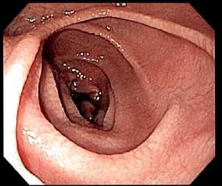
 Endoscopic still of duodenum of patient with coeliac disease showing scalloping of folds and "cracked-mud" appearance to mucosa
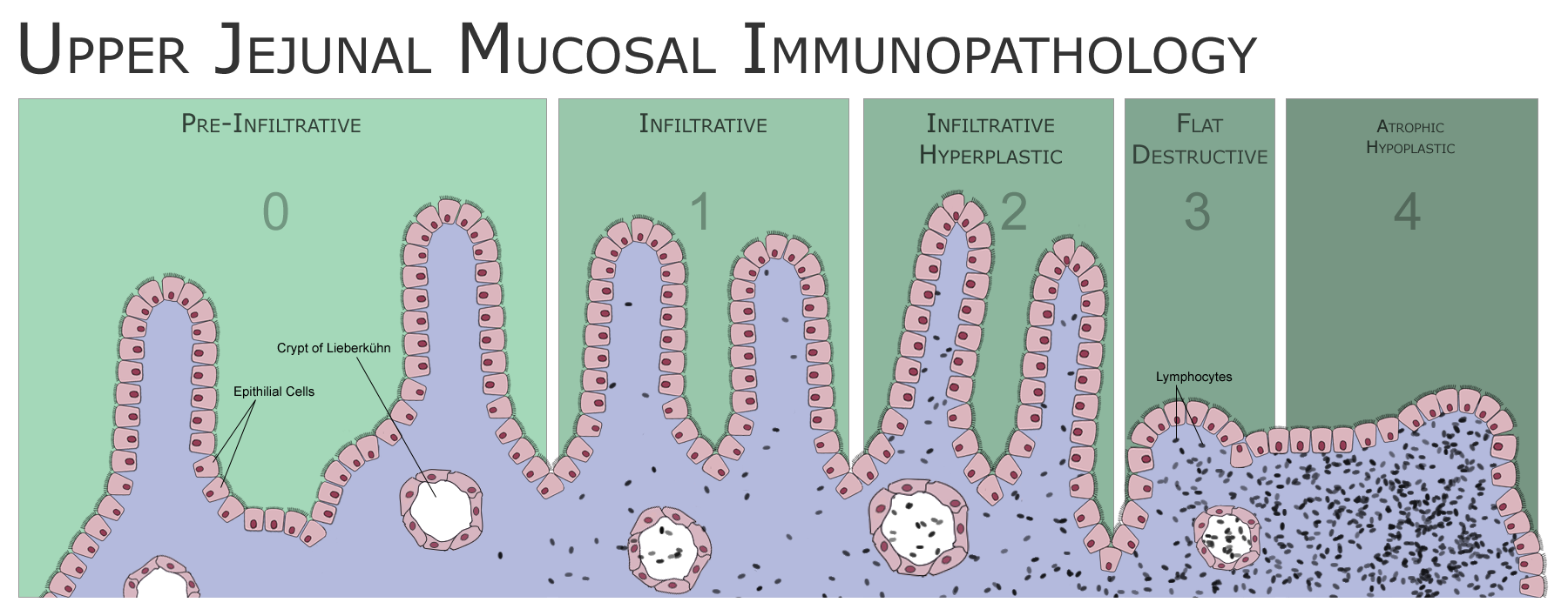
 Schematic of the Marsh classification of upper jejunal pathology in coeliac disease
An upper endoscopy with biopsy of the duodenum (beyond the duodenal bulb) or jejunum is performed. It is important for the physician to obtain multiple samples (four to eight) from the duodenum. Not all areas may be equally affected; if biopsies are taken from healthy bowel tissue, the result would be a false negative.[9]
Most patients with coeliac disease have a small bowel that appears normal on endoscopy; however, five concurrent endoscopic findings have been associated with a high specificity for coeliac disease: scalloping of the small bowel folds (pictured), paucity in the folds, a mosaic pattern to the mucosa (described as a "cracked-mud" appearance), prominence of the submucosa blood vessels, and a nodular pattern to the mucosa.[60]
Until the 1970s, biopsies were obtained using metal capsules attached to a suction device. The capsule was swallowed and allowed to pass into the small intestine. After x-ray verification of its position, suction was applied to collect part of the intestinal wall inside the capsule. Often-utilised capsule systems were the Watson capsule and the Crosby-Kugler capsule. This method has now been largely replaced by fibre-optic endoscopy, which carries a higher sensitivity and a lower frequency of errors.[61]
Pathology
The classic pathology changes of coeliac disease in the small bowel are categorised by the "Marsh classification":[62]
- Marsh stage 0: normal mucosa
- Marsh stage 1: increased number of intra-epithelial lymphocytes, usually exceeding 20 per 100 enterocytes
- Marsh stage 2: proliferation of the crypts of Lieberkuhn
- Marsh stage 3: partial or complete villous atrophy
- Marsh stage 4: hypoplasia of the small bowel architecture
Marsh's classification, introduced in 1992, was subsequently modified in 1999 to six stages, where the previous stage 3 was split in three substages.[63] Further studies demonstrated that this system was not always reliable and that the changes observed in coeliac disease could be described in one of three stages—A, B1 and B2—with A representing lymphocytic infiltration with normal villous appearance and B1 and B2 describing partial and complete villous atrophy.[4][64]
The changes classically improve or reverse after gluten is removed from the diet. However, most guidelines do not recommend a repeat biopsy unless there is no improvement in the symptoms on diet.[9][56] In some cases, a deliberate gluten challenge, followed by biopsy, may be conducted to confirm or refute the diagnosis. A normal biopsy and normal serology after challenge indicates the diagnosis may have been incorrect.[9]
Other diagnostic tests
At the time of diagnosis, further investigations may be performed to identify complications, such as iron deficiency (by full blood count and iron studies), folic acid and vitamin B12 deficiency and hypocalcaemia (low calcium levels, often due to decreased vitamin D levels). Thyroid function tests may be requested during blood tests to identify hypothyroidism, which is more common in people with coeliac disease.[10]
Osteopenia and osteoporosis, mildly and severely reduced bone mineral density, are often present in people with coeliac disease, and investigations to measure bone density may be performed at diagnosis, such as dual energy X-ray absorptiometry (DXA) scanning, to identify risk of fracture and need for bone protection medication.[9][10]
Screening
Due to its high sensitivity, serology has been proposed as a screening measure, because the presence of antibodies would detect previously undiagnosed cases of coeliac disease and prevent its complications in those patients.[9] There is significant debate as to the benefits of screening. Some studies suggest that early detection would decrease the risk of osteoporosis and anaemia. In contrast, a cohort study in Cambridge suggested that people with undetected coeliac disease had a beneficial risk profile for cardiovascular disease (less overweight, lower cholesterol levels).[2] There is limited evidence that screen-detected cases benefit from a diagnosis in terms of morbidity and mortality; hence, population-level screening is not presently thought to be beneficial.[4]
In the United Kingdom, the National Institute for Health and Clinical Excellence (NICE) recommends screening for coeliac disease in patients with newly diagnosed chronic fatigue syndrome[65] and irritable bowel syndrome,[8] as well as in type 1 diabetics, especially those with insufficient weight gain or unexplained weight loss.[17][66] It is also recommended in autoimmune thyroid disease, dermatitis herpetiformis, and in the first-degree relatives of those with confirmed coeliac disease.[17]
There is a large number of scenarios where testing for coeliac disease may be offered given previously described associations, such as the conditions mentioned above in "miscellaneous".[4][17]
Treatment
Diet
Main article: Gluten-free dietAt present, the only effective treatment is a life-long gluten-free diet.[19] No medication exists that will prevent damage or prevent the body from attacking the gut when gluten is present. Strict adherence to the diet allows the intestines to heal, leading to resolution of all symptoms in most cases and, depending on how soon the diet is begun, can also eliminate the heightened risk of osteoporosis and intestinal cancer and in some cases sterility.[67] Dietitian input is generally requested to ensure the patient is aware which foods contain gluten, which foods are safe, and how to have a balanced diet despite the limitations. In many countries, gluten-free products are available on prescription and may be reimbursed by health insurance plans.
The diet can be cumbersome; failure to comply with the diet may cause relapse. The term gluten-free is generally used to indicate a supposed harmless level of gluten rather than a complete absence.[68] The exact level at which gluten is harmless is uncertain and controversial. A recent systematic review tentatively concluded that consumption of less than 10 mg of gluten per day is unlikely to cause histological abnormalities, although it noted that few reliable studies had been done.[68] Regulation of the label gluten-free varies widely by country. In the United States, the FDA issued regulations in 2007 limiting the use of "gluten-free" in food products to those with less than 20 ppm of gluten.[69][70] The current international Codex Alimentarius standard allows for 20 ppm of gluten in so-called "gluten-free" foods.[71] Gluten-free products are usually more expensive and harder to find than common gluten-containing foods.[72] Since ready-made products often contain traces of gluten, some coeliacs may find it necessary to cook from scratch.[73]
Even while on a diet, health-related quality of life (HRQOL) may be lower in people with coeliac disease. Studies in the United States have found that quality of life becomes comparable to the general population after staying on the diet, while studies in Europe have found that quality of life remains lower, although the surveys are not quite the same.[74] Men tend to report more improvement than women.[75] Some have persisting digestive symptoms or dermatitis herpetiformis, mouth ulcers, osteoporosis and resultant fractures. Symptoms suggestive of irritable bowel syndrome may be present, and there is an increased rate of anxiety, fatigue, dyspepsia and musculoskeletal pain.[76]
Refractory disease
A tiny minority of patients suffer from refractory disease, which means they do not improve on a gluten-free diet. This may be because the disease has been present for so long that the intestines are no longer able to heal on diet alone, or because the patient is not adhering to the diet, or because the patient is consuming foods that are inadvertently contaminated with gluten. If alternative causes have been eliminated, steroids or immunosuppressants (such as azathioprine) may be considered in this scenario.[9]
Experimental treatments
Various other approaches are being studied that would reduce the need of dieting. All are still under development, and are not expected to be available to the general public for a while:[2]
- Genetically engineered wheat species, or wheat species that have been selectively bred to be minimally immunogenic. This, however, could interfere with the effects that gliadin has on the quality of dough.
- A combination of enzymes (prolyl endopeptidase and a barley glutamine-specific cysteine endopeptidase (EP-B2)) that degrade the putative 33-mer peptide in the duodenum. This combination would enable coeliac disease patients to consume gluten-containing products.[77]
- Inhibition of zonulin, an endogenous signalling protein linked to increased permeability of the bowel wall and hence increased presentation of gliadin to the immune system.[78]
- Scavenging of gliadin in the gastrointestinal tract by a polymeric binder to prevent its digestion into the immunogenic peptide.[79]
- Other treatments aimed at other well-understood steps in the pathogenesis of coeliac disease, such as the action of HLA-DQ2 or tissue transglutaminase and the MICA/NKG2D interaction that may be involved in the killing of enterocytes (bowel lining cells).
Epidemiology
The disease is thought to affect between 1 in 1750 (with CD defined as clinical cases including dermatitis herpetiformis) to 1 in 105 (CD defined by presence of IgA TG in blood donors) people in the United States.[3] The prevalence of clinically diagnosed disease (symptoms prompting diagnostic testing) is 0.05–0.27% in various studies. However, population studies from parts of Europe, India, South America, Australasia and the USA (using serology and biopsy) indicate that the prevalence may be between 0.33 and 1.06% in children (5.66% in one study of Sahrawi children[80]) and 0.18–1.2% in adults.[2] People of African, Japanese and Chinese descent are rarely diagnosed;[citation needed] this reflects a much lower prevalence of the genetic risk factors. Population studies also indicate that a large proportion of coeliacs remain undiagnosed; this is due, in part, to many clinicians being unfamiliar with the condition.[81]
Coeliac disease is more prevalent in women than in men.[82]
A large multicentre study in the U.S. found a prevalence of 0.75% in not-at-risk groups, rising to 1.8% in symptomatic patients, 2.6% in second-degree relatives of a patient with coeliac disease and 4.5% in first-degree relatives. This profile is similar to the prevalence in Europe.[83] Other populations at increased risk for coeliac disease, with prevalence rates ranging from 5% to 10%, include individuals with Down and Turner syndromes, type 1 diabetes, and autoimmune thyroid disease, including both hyperthyroidism (overactive thyroid) and hypothyroidism (underactive thyroid).[84]
Historically, coeliac disease was thought to be rare, with a prevalence of about 0.02%.[84] Recent increases in the number of reported cases may be due to changes in diagnostic practice,[85] but there is evidence that coeliac disease may be becoming more common in the United States.[86]
Social and religious issues
Christian churches & the Eucharist
Speaking generally, the various denominations of Christians celebrate a Eucharist in which a wafer or small piece of wheat bread is blessed and then eaten (see Sacramental bread). A typical wafer weighs about half a gram[87] Wheat flour contains around 10 to 13% gluten, so a single communion wafer may have more than 50 mg of gluten, an amount which will harm the health of many coeliac patients especially if consumed every day (see Diet above). Many Christian churches offer their communicants gluten-free alternatives, usually in the form of a rice-based cracker or gluten-free bread. These include United Methodist, Christian Reformed, Episcopal, Lutheran, and The Church of Jesus Christ of Latter-day Saints.[88]
Roman Catholic position
Roman Catholic doctrine states that for a valid Eucharist, the bread to be used at Mass must be made from wheat. In 2002, the Congregation for the Doctrine of the Faith approved German-made low-gluten hosts, which meets all of the Catholic Church's requirements, for use in Italy; although not entirely gluten-free, they were also approved by the Italian Celiac Association.[89] Some Catholic coeliac sufferers have requested permission to use rice wafers; such petitions have always been denied.[90] The issue is more complex for priests. Though a Catholic (lay or ordained) receiving the Eucharist under either form (bread or wine) is receiving Christ "whole and entire"—his body, blood, soul, and divinity—the priest, who is acting in persona Christi, is required to receive under both species when offering Mass—not for the validity of his Communion, but for the fullness of the sacrifice of the Mass. On 24 July 2003, the Congregation for the Doctrine of the Faith stated, "Given the centrality of the celebration of the Eucharist in the life of a priest, one must proceed with great caution before admitting to Holy Orders those candidates unable to ingest gluten or alcohol without serious harm."[91]
As of January 2004, an extremely low-gluten host became available in the United States. The Benedictine Sisters of Perpetual Adoration in Clyde, Missouri, produce low-gluten hosts safe for coeliacs and also approved by the Catholic Church for use at Mass. The hosts are made and packaged in a dedicated wheat-free, gluten-free environment. Gluten-content analysis found no detectable amount of gluten, though the reported gluten content is 0.01% as that was the lowest limit of detection possible with the utilised analysis technique. In an article from the Catholic Review (15 February 2004), Dr. Alessio Fasano was quoted as declaring these hosts "perfectly safe for celiac sufferers."[92]
Passover
The Jewish festival of Pesach (Passover) may present problems with its obligation to eat matzo, which is unleavened bread made in a strictly controlled manner from wheat, barley, spelt, oats, or rye. This rules out many other grains that are normally used as substitutes for people with gluten sensitivity, especially for Ashkenazi Jews, who also avoid rice. Many kosher-for-Passover products avoid grains altogether and are therefore gluten-free. Potato starch is the primary starch used to replace the grains. Consuming matzo is mandatory on the first night of Pesach only. Jewish law holds that a person should not seriously endanger one's health in order to fulfil a commandment. Thus, a person with severe coeliac disease is not required, or even allowed, to eat any matzo other than gluten-free matzo. The most commonly used gluten-free matzo is made from oats.[93]
History
Humans first started to cultivate grains in the Neolithic period (beginning about 9500 BCE) in the Fertile Crescent in Western Asia, and it is likely that coeliac disease did not occur before this time. Aretaeus of Cappadocia, living in the second century in the same area, recorded a malabsorptive syndrome with chronic diarrhoea. His "Cœliac Affection" (coeliac from Greek κοιλιακός koiliakos, "abdominal") gained the attention of Western medicine when Francis Adams presented a translation of Aretaeus's work at the Sydenham Society in 1856. The patient described in Aretaeus' work had stomach pain and was atrophied, pale, feeble and incapable of work. The diarrhoea manifested as loose stools that were white, malodorous and flatulent, and the disease was intractable and liable to periodic return. The problem, Aretaeus believed, was a lack of heat in the stomach necessary to digest the food and a reduced ability to distribute the digestive products throughout the body, this incomplete digestion resulting in the diarrhoea. He regarded this as an affliction of the old and more commonly affecting women, explicitly excluding children. The cause, according to Aretaeus, was sometimes either another chronic disease or even consuming "a copious draught of cold water."[5][6]
The paediatrician Samuel Gee gave the first modern-day description of the condition in children in a lecture at Hospital for Sick Children, Great Ormond Street, London, in 1887. Gee acknowledged earlier descriptions and terms for the disease and adopted the same term as Aretaeus (coeliac disease). He perceptively stated: "If the patient can be cured at all, it must be by means of diet." Gee recognised that milk intolerance is a problem with coeliac children and that highly starched foods should be avoided. However, he forbade rice, sago, fruit and vegetables, which all would have been safe to eat, and he recommended raw meat as well as thin slices of toasted bread. Gee highlighted particular success with a child "who was fed upon a quart of the best Dutch mussels daily." However, the child could not bear this diet for more than one season.[6][94]
Christian Archibald Herter, an American physician, wrote a book in 1908 on children with coeliac disease, which he called "intestinal infantilism." He noted their growth was retarded and that fat was better tolerated than carbohydrate. The eponym Gee-Herter disease was sometimes used to acknowledge both contributions.[95][96] Sidney V. Haas, an American paediatrician, reported positive effects of a diet of bananas in 1924.[97] This diet remained in vogue until the actual cause of coeliac disease was determined.[6]
While a role for carbohydrates had been suspected, the link with wheat was not made until the 1940s by the Dutch paediatrician Dr. Willem Karel Dicke.[98] It is likely that clinical improvement of his patients during the Dutch famine of 1944 (during which flour was scarce) may have contributed to his discovery.[99] Dicke noticed that the shortage of bread led to a significant drop in the death rate among children affected by CD from greater than 35% to essentially zero. He also reported that once wheat was again available after the conflict, the mortality rate soared to previous levels.[100] The link with the gluten component of wheat was made in 1952 by a team from Birmingham, England.[101] Villous atrophy was described by British physician John W. Paulley in 1954 on samples taken at surgery.[102] This paved the way for biopsy samples taken by endoscopy.[6]
Throughout the 1960s, other features of coeliac disease were elucidated. Its hereditary character was recognised in 1965.[103] In 1966, dermatitis herpetiformis was linked to gluten sensitivity.[6][13]
May has been designated as "Coeliac Awareness Month".[104][105]
References
- ^ See American and British English spelling differences.
- ^ a b c d e f g h i j k l m n van Heel D, West J (2006). "Recent advances in coeliac disease". Gut 55 (7): 1037–46. doi:10.1136/gut.2005.075119. PMC 1856316. PMID 16766754. http://gut.bmjjournals.com/cgi/content/full/55/7/1037.
- ^ a b Rewers, Marian J. (2005). "Epidemiology of Celiac Disease: What Are the Prevalence, Incidence, and Progression of Celiac Disease?". Gastroenterology (National Institute of Health) 128 (4 Suppl 1): S47–51. doi:10.1053/j.gastro.2005.02.030. PMID 15825126. http://consensus.nih.gov/2004/2004CeliacDisease118Program.pdf#page=40
- ^ a b c d e f g h i j k l Di Sabatino A, Corazza GR (April 2009). "Coeliac disease". Lancet 373 (9673): 1480–93. doi:10.1016/S0140-6736(09)60254-3. PMID 19394538.
- ^ a b Adams F, translator (1856). "On The Cœliac Affection". The extant works of Aretaeus, The Cappadocian. London: Sydenham Society. pp. 350–1. http://books.google.com/books?id=v4gIAAAAIAAJ&pg=PA350#v=onepage&q=&f=false. Retrieved 12 December 2009.
- ^ a b c d e f Losowsky MS (2008). "A history of coeliac disease". Dig Dis 26 (2): 112–20. doi:10.1159/000116768. PMID 18431060.
- ^ Ferguson R, Basu M, Asquith P, Cooke W (1976). "Jejunal mucosal abnormalities in patients with recurrent aphthous ulceration". Br Med J 1 (6000): 11–13. doi:10.1136/bmj.1.6000.11. PMC 1638254. PMID 1247715. http://www.pubmedcentral.nih.gov/articlerender.fcgi?tool=pmcentrez&artid=1638254.
- ^ a b National Institute for Health and Clinical Excellence. Clinical guideline 61: Irritable bowel syndrome. London, 2008.
- ^ a b c d e f g h i "American Gastroenterological Association medical position statement: Celiac Sprue". Gastroenterology 120 (6): 1522–5. 2001. doi:10.1053/gast.2001.24055. PMID 11313323. http://www.gastrojournal.org/article/PIIS0016508501701618/fulltext.
- ^ a b c Presutti RJ, Cangemi JR, Cassidy HD, Hill DA (December 2007). "Celiac disease". Am Fam Physician 76 (12): 1795–802. PMID 18217518. http://www.aafp.org/afp/20071215/1795.html.
- ^ Tursi A, Brandimarte G, Giorgetti G (2003). "High prevalence of small intestinal bacterial overgrowth in celiac patients with persistence of gastrointestinal symptoms after gluten withdrawal". Am J Gastroenterol 98 (4): 839–43. doi:10.1111/j.1572-0241.2003.07379.x. PMID 12738465.
- ^ Cunningham-Rundles C (September 2001). "Physiology of IgA and IgA deficiency". J. Clin. Immunol. 21 (5): 303–9. doi:10.1023/A:1012241117984. PMID 11720003.
- ^ a b Marks J, Shuster S, Watson A (1966). "Small-bowel changes in dermatitis herpetiformis". Lancet 2 (7476): 1280–2. doi:10.1016/S0140-6736(66)91692-8. PMID 4163419.
- ^ Nicolas ME, Krause PK, Gibson LE, Murray JA (August 2003). "Dermatitis herpetiformis". Int. J. Dermatol. 42 (8): 588–600. doi:10.1046/j.1365-4362.2003.01804.x. PMID 12890100.
- ^ Ferguson A, Hutton M, Maxwell J, Murray D (1970). "Adult coeliac disease in hyposplenic patients". Lancet 1 (7639): 163–4. doi:10.1016/S0140-6736(70)90405-8. PMID 4189238.
- ^ Collin P, Kaukinen K, Välimäki M, Salmi J (2002). "Endocrinological disorders and celiac disease". Endocr Rev 23 (4): 464–83. doi:10.1210/er.2001-0035. PMID 12202461. http://edrv.endojournals.org/cgi/content/full/23/4/464.
- ^ a b c d e f g h i National Institute for Health and Clinical Excellence. Clinical guideline 86: Recognition and assessment of coeliac disease. London, 2009.
- ^ Schuppan D, Junker Y, Barisani D (December 2009). "Celiac disease: from pathogenesis to novel therapies". Gastroenterology 137 (6): 1912–33. doi:10.1053/j.gastro.2009.09.008. PMID 19766641. http://www.gastrojournal.org/article/PIIS001650850901600X/fulltext.
- ^ a b c d e Kupper C (2005). "Dietary guidelines and implementation for celiac disease". Gastroenterology 128 (4 Suppl 1): S121–7. doi:10.1053/j.gastro.2005.02.024. PMID 15825119.
- ^ Gallagher, Eimear (2009). Gluten-free Food Science and Technology. Published by John Wiley and Sons,. pp. 320. ISBN 1405159154, 9781405159159. http://eu.wiley.com/WileyCDA/WileyTitle/productCd-1405159154.html.
- ^ a b c d Hadithi M, von Blomberg BM, Crusius JB, et al. (2007). "Accuracy of serologic tests and HLA-DQ typing for diagnosing celiac disease". Ann. Intern. Med. 147 (5): 294–302. PMID 17785484. http://www.annals.org/cgi/content/full/147/5/294.
- ^ Kim C, Quarsten H, Bergseng E, Khosla C, Sollid L (2004). "Structural basis for HLA-DQ2-mediated presentation of gluten epitopes in celiac disease". Proc Natl Acad Sci USA 101 (12): 4175–9. doi:10.1073/pnas.0306885101. PMC 384714. PMID 15020763. http://www.pnas.org/cgi/content/full/101/12/4175.
- ^ Jores RD, Frau F, Cucca F, et al. (2007). "HLA-DQB1*0201 homozygosis predisposes to severe intestinal damage in celiac disease". Scand. J. Gastroenterol. 42 (1): 48–53. doi:10.1080/00365520600789859. PMID 17190762.
- ^ Karell K, Louka AS, Moodie SJ, et al. (2003). "HLA types in celiac disease patients not carrying the DQA1*05-DQB1*02 (DQ2) heterodimer: results from the European Genetics Cluster on Celiac Disease". Hum. Immunol. 64 (4): 469–77. doi:10.1016/S0198-8859(03)00027-2. PMID 12651074.
- ^ Michalski J, McCombs C, Arai T, Elston R, Cao T, McCarthy C, Stevens F (1996). "HLA-DR, DQ genotypes of celiac disease patients and healthy subjects from the West of Ireland". Tissue Antigens 47 (2): 127–33. doi:10.1111/j.1399-0039.1996.tb02525.x. PMID 8851726.
- ^ Kaur G, Sarkar N, Bhatnagar S, et al. (2002). "Pediatric celiac disease in India is associated with multiple DR3-DQ2 haplotypes". Hum. Immunol. 63 (8): 677–82. doi:10.1016/S0198-8859(02)00413-5. PMID 12121676.
- ^ Layrisse Z, Guedez Y, Domínguez E, Paz N, Montagnani S, Matos M, Herrera F, Ogando V, Balbas O, Rodríguez-Larralde A (2001). "Extended HLA haplotypes in a Carib Amerindian population: the Yucpa of the Perija Range". Hum Immunol 62 (9): 992–1000. doi:10.1016/S0198-8859(01)00297-X. PMID 11543901.
- ^ a b Dubois et al.; Trynka, G; Franke, L; Hunt, KA; Romanos, J; Curtotti, A; Zhernakova, A; Heap, GA et al. (2010). "Multipel common variants for celiac diease influencing immune gene expression". Nature Genetics 42 (4): 295–302. doi:10.1038/ng.543. PMC 2847618. PMID 20190752. http://www.pubmedcentral.nih.gov/articlerender.fcgi?tool=pmcentrez&artid=2847618.
- ^ Dwain N. Walcher, Norman Kretchmer. Food, nutrition, and evolution: food as an environmental factor in the genesis of human variability. pp. 179-199.
- ^ Catassi, Carlo (2005). "Where Is Celiac Disease Coming From and Why?". Journal of Pediatric Gastroenterology & Nutrition. http://journals.lww.com/jpgn/Fulltext/2005/03000/Where_Is_Celiac_Disease_Coming_From_and_Why_.6.aspx.
- ^ Zhernakova A et al (2010). "Evolutionary and Functional Analysis of Celiac Risk Loci Reveals SH2B3 as a Protective Factor against Bacterial Infection". The American Journal of Human Genetics.
- ^ Green PH, Cellier C (October 2007). "Celiac disease". N. Engl. J. Med. 357 (17): 1731–43. doi:10.1056/NEJMra071600. PMID 17960014.
- ^ Lammers KM, Lu R, Brownley J, et al. (July 2008). "Gliadin induces an increase in intestinal permeability and zonulin release by binding to the chemokine receptor CXCR3". Gastroenterology 135 (1): 194–204.e3. doi:10.1053/j.gastro.2008.03.023. PMC 2653457. PMID 18485912. http://www.pubmedcentral.nih.gov/articlerender.fcgi?tool=pmcentrez&artid=2653457.
- ^ a b Qiao SW, Bergseng E, Molberg Ø, et al. (August 2004). "Antigen presentation to celiac lesion-derived T cells of a 33-mer gliadin peptide naturally formed by gastrointestinal digestion". J. Immunol. 173 (3): 1757–62. PMID 15265905. http://www.jimmunol.org/cgi/pmidlookup?view=long&pmid=15265905.
- ^ Shan L, Qiao SW, Arentz-Hansen H, et al. (2005). "Identification and analysis of multivalent proteolytically resistant peptides from gluten: implications for celiac sprue". J. Proteome Res. 4 (5): 1732–41. doi:10.1021/pr050173t. PMC 1343496. PMID 16212427. http://www.pubmedcentral.nih.gov/articlerender.fcgi?tool=pmcentrez&artid=1343496.
- ^ Dieterich W, Ehnis T, Bauer M, Donner P, Volta U, Riecken E, Schuppan D (1997). "Identification of tissue transglutaminase as the autoantigen of celiac disease". Nat Med 3 (7): 797–801. doi:10.1038/nm0797-797. PMID 9212111.
- ^ Skovbjerg H, Norén O, Anthonsen D, Moller J, Sjöström H (2002). "Gliadin is a good substrate of several transglutaminases: possible implication in the pathogenesis of coeliac disease.". Scand J Gastroenterol 37 (7): 812–7. doi:10.1080/713786534. PMID 12190095.
- ^ Fleckenstein B, Molberg 0, Qiao SW, Schmid DG, Mulbe E von der, Elgstoen K, Jung G and Soflid ML. (2002). "Gliadin T cell epitope selection by tissue transglutaminase in celiac disease. Role of enzyme specificity and pH influence on the transamidation versus deamidation process.". J Biol Chem 277 (37): 34109–34116. doi:10.1074/jbc.M204521200. PMID 12093810.
- ^ Koning, F. .; Schuppan, D. .; Cerf-Bensussan, N. .; Sollid, M. . (Jun 2005). "Pathomechanisms in celiac disease". Best practice & research. Clinical gastroenterology 19 (3): 373–387. doi:10.1016/j.bpg.2005.02.003. ISSN 1521-6918. PMID 15925843.
- ^ Mowat AM. (2003). "Coeliac disease – a meeting point for genetics, immunology, and protein chemistry". Lancet 361 (9365): 1290–1292. doi:10.1016/S0140-6736(03)12989-3. PMID 12699968.
- ^ Dewar D, Pereira SP and Ciclitira PJ (2004). "The pathogenesis of coeliac disease". Int J Biochem Cell Biol 36 (1): 17–24. doi:10.1016/S1357-2725(03)00239-5. PMID 14592529.
- ^ Kaukinen K, Peraaho M, Collin P, Partanen J, Woolley N, Kaartinen T, Nuuntinen T, Halttunen T, Maki M, Korponay-Szabo I (2005). "Small-bowel mucosal tranglutaminase 2-specific IgA deposits in coeliac disease without villous atrophy: A Prospective and radmonized clinical study". Scand J Gastroenterology 40 (5): 564–572. doi:10.1080/00365520510023422. PMID 16036509.
- ^ Salmi T, Collin P, Korponay-Szabó I, Laurila K, Partanen J, Huhtala H, Király R, Lorand L, Reunala T, Mäki M, Kaukinen K (2006). "Endomysial antibody-negative coeliac disease: clinical characteristics and intestinal autoantibody deposits". Gut 55 (12): 1746–53. doi:10.1136/gut.2005.071514. PMC 1856451. PMID 16571636. http://gut.bmj.com/cgi/content/full/55/12/1746.
- ^ Londei M, Ciacci C, Ricciardelli I, Vacca L, Quaratino S, and Maiuri L. (2005). "Gliadin as a stimulator of innate responses in celiac disease". Mol Immunol 42 (8): 913–918. doi:10.1016/j.molimm.2004.12.005. PMID 15829281.
- ^ Zanoni G, Navone R, Lunardi C, Tridente G, Bason C, Sivori S, Beri R, Dolcino M, Valletta E, Corrocher R, Puccetti A (2006). "In celiac disease, a subset of autoantibodies against transglutaminase binds toll-like receptor 4 and induces activation of monocytes". PLoS Med 3 (9): e358. doi:10.1371/journal.pmed.0030358. PMC 1569884. PMID 16984219. http://medicine.plosjournals.org/perlserv/?request=get-document&doi=10.1371/journal.pmed.0030358.
- ^ Salim A, Phillips A, Farthing M (1990). "Pathogenesis of gut virus infection". Baillieres Clin Gastroenterol 4 (3): 593–607. doi:10.1016/0950-3528(90)90051-H. PMID 1962725.
- ^ Stene L, Honeyman M, Hoffenberg E, Haas J, Sokol R, Emery L, Taki I, Norris J, Erlich H, Eisenbarth G, Rewers M (2006). "Rotavirus infection frequency and risk of celiac disease autoimmunity in early childhood: a longitudinal study". Am J Gastroenterol 101 (10): 2333–40. doi:10.1111/j.1572-0241.2006.00741.x. PMID 17032199.
- ^ Kagnoff M, Paterson Y, Kumar P, Kasarda D, Carbone F, Unsworth D, Austin R (1987). "Evidence for the role of a human intestinal adenovirus in the pathogenesis of coeliac disease" (PDF). Gut 28 (8): 995–1001. doi:10.1136/gut.28.8.995. PMC 1433141. PMID 2822550. http://gut.bmj.com/cgi/reprint/28/8/995.
- ^ Suman S, Williams E, Thomas P, Surgenor S, Snook J (2003). "Is the risk of adult coeliac disease causally related to cigarette exposure?". Eur J Gastroenterol Hepatol 15 (9): 995–1000. doi:10.1097/00042737-200309000-00009. PMID 12923372.
- ^ Norris JM, Barriga K, Hoffenberg EJ, Taki I, Miao D, Haas JE, Emery LM, Sokol RJ, Erlich HA, Eisenbarth GS, Rewers M. (2005). "Risk of celiac disease autoimmunity and timing of gluten introduction in the diet of infants at increased risk of disease". JAMA 293 (19): 2343–2351. doi:10.1001/jama.293.19.2343. PMID 15900004. http://jama.ama-assn.org/cgi/content/full/293/19/2343.
- ^ Poole J, Barriga K, Leung D, Hoffman M, Eisenbarth G, Rewers M, Norris J (2006). "Timing of initial exposure to cereal grains and the risk of wheat allergy". Pediatrics 117 (6): 2175–82. doi:10.1542/peds.2005-1803. PMID 16740862. http://pediatrics.aappublications.org/cgi/content/full/117/6/2175.
- ^ Akobeng A, Ramanan A, Buchan I, Heller R (2006). "Effect of breast feeding on risk of coeliac disease: a systematic review and meta-analysis of observational studies". Arch Dis Child 91 (1): 39–43. doi:10.1136/adc.2005.082016. PMC 2083075. PMID 16287899. http://adc.bmj.com/cgi/content/full/91/1/39.
- ^ Hopper A, Cross S, Hurlstone D, McAlindon M, Lobo A, Hadjivassiliou M, Sloan M, Dixon S, Sanders D (2007). "Pre-endoscopy serological testing for coeliac disease: evaluation of a clinical decision tool". BMJ 334 (7596): 729. doi:10.1136/bmj.39133.668681.BE. PMC 1847864. PMID 17383983. http://www.bmj.com/cgi/content/full/334/7596/729.
- ^ a b van der Windt DA, Jellema P, Mulder CJ, Kneepkens CM, van der Horst HE (2010). "Diagnostic testing for celiac disease among patients with abdominal symptoms: a systematic review.". JAMA 303 (17): 1738–46. doi:10.1001/jama.2010.549. PMID 20442390.
- ^ Sblattero D, Berti I, Trevisiol C, et al. (May 2000). "Human recombinant tissue transglutaminase ELISA: an innovative diagnostic assay for celiac disease". Am. J. Gastroenterol. 95 (5): 1253–7. doi:10.1111/j.1572-0241.2000.02018.x. PMID 10811336. http://www3.interscience.wiley.com/cgi-bin/fulltext/119001630/HTMLSTART.
- ^ a b Hill ID, Dirks MH, Liptak GS, et al. (January 2005). "Guideline for the diagnosis and treatment of celiac disease in children: recommendations of the North American Society for Pediatric Gastroenterology, Hepatology and Nutrition". J. Pediatr. Gastroenterol. Nutr. 40 (1): 1–19. doi:10.1097/00005176-200501000-00001. PMID 15625418. http://journals.lww.com/jpgn/Fulltext/2005/01000/Guideline_for_the_Diagnosis_and_Treatment_of.1.aspx.
- ^ Hill ID (April 2005). "What are the sensitivity and specificity of serologic tests for celiac disease? Do sensitivity and specificity vary in different populations?". Gastroenterology 128 (4 Suppl 1): S25–32. doi:10.1053/j.gastro.2005.02.012. PMID 15825123. http://consensus.nih.gov/2004/2004CeliacDisease118Program.pdf.
- ^ Wong R, Steele R, Reeves G, Wilson R, Pink A, Adelstein S (2003). "Antibody and genetic testing in coeliac disease". Pathology 35 (4): 285–304. PMID 12959764.
- ^ Korponay-Szabó I, Dahlbom I, Laurila K, Koskinen S, Woolley N, Partanen J, Kovács J, Mäki M, Hansson T (2003). "Elevation of IgG antibodies against tissue transglutaminase as a diagnostic tool for coeliac disease in selective IgA deficiency". Gut 52 (11): 1567–71. doi:10.1136/gut.52.11.1567. PMC 1773847. PMID 14570724. http://gut.bmjjournals.com/cgi/content/full/52/11/1567.
- ^ Niveloni S, Fiorini A, Dezi R, Pedreira S, Smecuol E, Vazquez H, Cabanne A, Boerr LA, Valero J, Kogan Z, Maurino E, Bai JC. (1998). "Usefulness of videoduodenoscopy and vital dye staining as indicators of mucosal atrophy of celiac disease: assessment of interobserver agreement". Gastrointestinal Endoscopy 47 (3): 223–229. doi:10.1016/S0016-5107(98)70317-7. PMID 9580349.
- ^ Mee A, Burke M, Vallon A, Newman J, Cotton P (1985). "Small bowel biopsy for malabsorption: comparison of the diagnostic adequacy of endoscopic forceps and capsule biopsy specimens". Br Med J (Clin Res Ed) 291 (6498): 769–72. doi:10.1136/bmj.291.6498.769. PMC 1417146. PMID 3929934. http://www.pubmedcentral.nih.gov/articlerender.fcgi?tool=pmcentrez&artid=1417146.
- ^ Marsh M (1992). "Gluten, major histocompatibility complex, and the small intestine. A molecular and immunobiologic approach to the spectrum of gluten sensitivity ('celiac sprue')". Gastroenterology 102 (1): 330–54. PMID 1727768.
- ^ Oberhuber G, Granditsch G, Vogelsang H (October 1999). "The histopathology of coeliac disease: time for a standardized report scheme for pathologists". Eur J Gastroenterol Hepatol 11 (10): 1185–94. doi:10.1097/00042737-199910000-00019. PMID 10524652.
- ^ Corazza GR, Villanacci V, Zambelli C, et al. (July 2007). "Comparison of the interobserver reproducibility with different histologic criteria used in celiac disease". Clin. Gastroenterol. Hepatol. 5 (7): 838–43. doi:10.1016/j.cgh.2007.03.019. PMID 17544877.
- ^ National Institute for Health and Clinical Excellence. Clinical guideline 53: Chronic fatigue syndrome/myalgic encephalomyelitis. London, 2007.
- ^ National Institute for Health and Clinical Excellence. Clinical guideline 15: Diagnosis and management of type 1 diabetes in children, young people and adults. London, 2004.
- ^ Treem W (2004). "Emerging concepts in celiac disease". Curr Opin Pediatr 16 (5): 552–9. doi:10.1097/01.mop.0000142347.74135.73. PMID 15367850.
- ^ a b Akobeng AK, Thomas AG (June 2008). "Systematic review: tolerable amount of gluten for people with coeliac disease". Aliment. Pharmacol. Ther. 27 (11): 1044–52. doi:10.1111/j.1365-2036.2008.03669.x. PMID 18315587.
- ^ Section 206 of the Food Allergen Labeling and Consumer Protection Act of 2004, Title II of Pub.L. 108-282, 118 Stat. 891, enacted August 2, 2004
- ^ 72 F.R. 2795-2817
- ^ "Current Official Standards". FAO/WHO. http://www.codexalimentarius.net/web/standard_list.jsp. Retrieved 26 June 2008.
- ^ Lee AR, Ng DL, Zivin J, Green PH (October 2007). "Economic burden of a gluten-free diet". J Hum Nutr Diet 20 (5): 423–30. doi:10.1111/j.1365-277X.2007.00763.x. PMID 17845376.
- ^ Troncone R, Ivarsson A, Szajewska H, Mearin ML (June 2008). "Review article: future research on coeliac disease – a position report from the European multistakeholder platform on coeliac disease (CDEUSSA)". Aliment. Pharmacol. Ther. 27 (11): 1030–43. doi:10.1111/j.1365-2036.2008.03668.x. PMID 18315588.
- ^ Häuser W, Stallmach A, Caspary WF, Stein J (March 2007). "Predictors of reduced health-related quality of life in adults with coeliac disease". Aliment. Pharmacol. Ther. 25 (5): 569–78. doi:10.1111/j.1365-2036.2006.03227.x. PMID 17305757.
- ^ Goddard CJ, Gillett HR (November 2006). "Complications of coeliac disease: are all patients at risk?". Postgrad Med J 82 (973): 705–12. doi:10.1136/pgmj.2006.048876. PMC 2660494. PMID 17099088. http://pmj.bmj.com/cgi/content/full/82/973/705.
- ^ Häuser W, Gold J, Stein J, Caspary WF, Stallmach A (July 2006). "Health-related quality of life in adult coeliac disease in Germany: results of a national survey". Eur J Gastroenterol Hepatol 18 (7): 747–54. doi:10.1097/01.meg.0000221855.19201.e8. PMID 16772832.
- ^ Siegel M, Bethune M, Gass J, Ehren J, Xia J, Johannsen A, Stuge T, Gray G, Lee P, Khosla C (2006). "Rational design of combination enzyme therapy for celiac sprue". Chem Biol 13 (6): 649–58. doi:10.1016/j.chembiol.2006.04.009. PMID 16793522. http://www.chembiol.com/content/article/fulltext?uid=PIIS1074552106001499.
- ^ Fasano A, Not T, Wang W, Uzzau S, Berti I, Tommasini A, Goldblum S (2000). "Zonulin, a newly discovered modulator of intestinal permeability, and its expression in coeliac disease". Lancet 355 (9214): 1518–9. doi:10.1016/S0140-6736(00)02169-3. PMID 10801176.
- ^ Pinier, M.; Verdu E.F.; Nasser-Eddine M.; David C.S.; Vézina A.; Rivard N. and Leroux J.C. (2009) Polymeric Binders Suppress Gliadin-Induced Toxicity in the Intestinal Epithelium. Gastroenterology, 136, 288-298 abstract
- ^ Catassi C, Rätsch I, Gandolfi L, Pratesi R, Fabiani E, El Asmar R, Frijia M, Bearzi I, Vizzoni L (1999). "Why is coeliac disease endemic in the people of the Sahara?". Lancet 354 (9179): 647–8. doi:10.1016/S0140-6736(99)02609-4. PMID 10466670.
- ^ Zipser R, Farid M, Baisch D, Patel B, Patel D (2005). "Physician awareness of celiac disease: a need for further education". J Gen Intern Med 20 (7): 644–6. doi:10.1007/s11606-005-0111-7. PMC 1490146. PMID 16050861. http://www3.interscience.wiley.com/journal/118700823/abstract.
- ^ Hischenhuber, C; Crevel, R; Jarry, B; Mäki, M; Moneret-Vautrin, D A; Romano, A; Troncone, R; Ward, R (March 2006). "Review article: safe amounts of gluten for patients with wheat allergy or coeliac disease". Aliment. Pharmacol. Ther. 23 (5): 559–75. doi:10.1111/j.1365-2036.2006.02768.x. PMID 16480395. http://onlinelibrary.wiley.com/doi/10.1111/j.1365-2036.2006.02768.x/full.
- ^ Fasano A, Berti I, Gerarduzzi T, Not T, Colletti R, Drago S, Elitsur Y, Green P, Guandalini S, Hill I, Pietzak M, Ventura A, Thorpe M, Kryszak D, Fornaroli F, Wasserman S, Murray J, Horvath K (2003). "Prevalence of celiac disease in at-risk and not-at-risk groups in the United States: a large multicenter study". Archives of Internal Medicine 163 (3): 286–92. doi:10.1001/archinte.163.3.286. PMID 12578508. http://archinte.ama-assn.org/cgi/content/full/163/3/286.
- ^ a b Barker JM, Liu E (2008). "Celiac disease: pathophysiology, clinical manifestations, and associated autoimmune conditions". Adv Pediatr 55: 349–65. doi:10.1016/j.yapd.2008.07.001. PMC 2775561. PMID 19048738. http://www.pubmedcentral.nih.gov/articlerender.fcgi?tool=pmcentrez&artid=2775561.
- ^ Leeds JS, Hopper AD, Sanders DS (2008). "Coeliac disease". Br Med Bull 88 (1): 157–70. doi:10.1093/bmb/ldn044. PMID 19073695.
- ^ Rubio-Tapia et al.; Kyle, RA; Kaplan, EL; Johnson, DR; Page, W; Erdtmann, F; Brantner, TL; Kim, WR et al. (2009). "Increased Prevalence and Mortality in Undiagnosed Celiac Disease". Gastroenterology 137 (1): 88–93. doi:10.1053/j.gastro.2009.03.059. PMC 2704247. PMID 19362553. http://www.gastrojournal.org/article/S0016-5085(09)00523-X/abstract.
- ^ One on-line site sells 1200 wafers weighing a total of 523 g
- ^ Jax Peter Lowell, The Gluten-Free Bible, p. 279.
- ^ Scott Adams (2 August 2002). "Bishops in Italy Approve a German-made Low Gluten Eucharistic Host". Celiac.com. http://www.celiac.com/st_prod.html?p_prodid=696.
- ^ Associated Press (8 December 2004). "Girl with digestive disease denied Communion". MSNBC (Microsoft). http://www.msnbc.msn.com/id/5762478/. Retrieved 30 May 2006.
- ^ Ratzinger, Joseph (24 July 2003). Prot. 89/78-174 98. Congregation for the Doctrine of the Faith. Full text at: "The Use of Mustum and Low-Gluten Hosts at Mass". BCL Newsletter. United States Conference of Catholic Bishops. November 2003. http://www.usccb.org/liturgy/innews/1103.shtml. Retrieved 7 March 2007.
- ^ McNamara, Father Edward (15 September 2004). "Liturgy: Gluten-free Hosts". Catholic Online. http://www.catholic.org/featured/headline.php?ID=1340. Retrieved 17 June 2007.
- ^ Rabbi Avraham Juravel. "Gluten Intolerance, Celiac, Allergies And Pesach". Orthodox Union. http://oukosher.org/index.php/articles/single/gluten_intolerance_celiac_allergies_and_pesach/. Retrieved 3 September 2006.
- ^ Gee, SJ (1888). "On the coeliac affection". St Bartholomew's Hospital Report 24: 17–20. http://web2.bium.univ-paris5.fr/livanc/?cote=epo0466&p=1&do=page.
- ^ Herter, CA (1908). On infantilism from chronic intestinal infection; characterized by the overgrowth and persistence of flora in the nursing period. New York: Macmillan & Co. as cited by WhoNamedIt
- ^ Ole Daniel Enersen. "Christian Archibald Herter". Who Named It?. http://www.whonamedit.com/doctor.cfm/1490.html. Retrieved 20 March 2007.
- ^ Haas SV (1924). "The value of the banana in the treatment of coeliac disease". Am J Dis Child 24: 421–37.
- ^ van Berge-Henegouwen G, Mulder C (1993). "Pioneer in the gluten free diet: Willem-Karel Dicke 1905–1962, over 50 years of gluten free diet" (PDF). Gut 34 (11): 1473–5. doi:10.1136/gut.34.11.1473. PMC 1374403. PMID 8244125. http://gut.bmj.com/cgi/reprint/34/11/1473.
- ^ Dicke WK (1950) (in Dutch). Coeliakie: een onderzoek naar de nadelige invloed van sommige graansoorten op de lijder aan coeliakie, PhD thesis. Utrecht, the Netherlands: University of Utrecht.
- ^ Fasano A (2009). "Celiac Disease Insights: Clues to Solving Autoimmunity". Scientific American (August): 49–57. http://www.scientificamerican.com/article.cfm?id=celiac-disease-insights.
- ^ Anderson C, French J, Sammons H, Frazer A, Gerrard J, Smellie J (1952). "Coeliac disease; gastrointestinal studies and the effect of dietary wheat flour". Lancet 1 (17): 836–42. doi:10.1016/S0140-6736(52)90795-2. PMID 14918439.
- ^ Paulley JW (1954). "Observation on the aetiology of idiopathic steatorrhoea; jejunal and lymph-node biopsies". Br Med J 4900 (4900): 1318–21. doi:10.1136/bmj.2.4900.1318. PMC 2080246. PMID 13209109. http://www.pubmedcentral.nih.gov/articlerender.fcgi?tool=pmcentrez&artid=2080246.
- ^ Macdonald W, Dobbins W, Rubin C (1965). "Studies of the familial nature of celiac sprue using biopsy of the small intestine". N Engl J Med 272 (9): 448–56. doi:10.1056/NEJM196503042720903. PMID 14242522.
- ^ "Buy Me Some Gluten-Free Peanuts, Cracker Jacks". QSR magazine (Journalistic). May 11, 2010. http://www.qsrmagazine.com/news/buy-me-some-gluten-free-peanuts-cracker-jacks. Retrieved December 30, 2010.
- ^ Hillson, Beth (January 9, 2008). "May as Celiac Awareness Month". Celiac Disease Foundation. http://www.celiac.org/index.php?option=com_content&view=article&id=86&Itemid=119. Retrieved July 1, 2011.
External links
- Coeliac disease at the Open Directory Project
- Celiac Disease Awareness Campaign from the National Institutes of Health
- Celiac Disease practice guideline from the World Gastroenterology Organisation (WGO)
- Outcomes of 2004 consensus development conference, U.S. National Institutes of Health
Immune disorders: hypersensitivity and autoimmune diseases (279.5–6) Type I/allergy/atopy
(IgE)ForeignAtopic dermatitis · Allergic urticaria · Hay fever · Allergic asthma · Anaphylaxis · Food allergy (Milk, Egg, Peanut, Tree nut, Seafood, Soy, Wheat), Penicillin allergyAutoimmunenoneType II/ADCC
(IgM, IgG)ForeignAutoimmuneAutoimmune hemolytic anemia · Idiopathic thrombocytopenic purpura · Bullous pemphigoid · Pemphigus vulgaris · Rheumatic fever · Goodpasture's syndromeType III
(Immune complex)ForeignHenoch–Schönlein purpura · Hypersensitivity vasculitis · Reactive arthritis · Rheumatoid arthritis · Farmer's lung · Post-streptococcal glomerulonephritis · Serum sickness · Arthus reactionAutoimmuneType IV/cell-mediated
(T-cells)ForeignAllergic contact dermatitis · Mantoux testAutoimmuneDiabetes mellitus type 1 · Hashimoto's thyroiditis · Guillain–Barré syndrome · Multiple sclerosis · Coeliac disease · Giant-cell arteritisUnknown/
multipleForeignAutoimmuneSjögren's syndrome · Autoimmune hepatitis · Autoimmune polyendocrine syndrome (APS1, APS2) · Autoimmune adrenalitis · Systemic autoimmune diseaseGluten sensitivity Conditions general: Wheat allergy · Oat sensitivity
nervous system: GS idiopathic neuropathies
digestive system: Coeliac disease · GSE associated conditions
integumentary system: Dermatitis herpetiformisAntibodies HLA-DQ Other Categories:- Autoimmune diseases
- GI tract disorders
- Genetic disorders by system
- Malnutrition
- Pediatrics


Wikimedia Foundation. 2010.