- Crus of diaphragm
-
Crus of diaphragm 
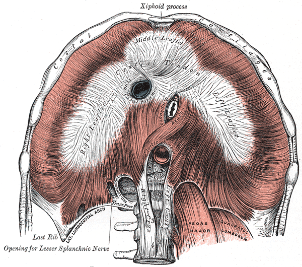
The diaphragm. Under surface. (Left crus and right crus are at bottom center.) 
Deep and superficial dissection of the lumbar plexus. (Rt. crus of diaph. labeled vertically at center.) Latin crus sinistrum diaphragmatis, crus dextrum diaphragmatis Gray's subject #117 405 The crura of the diaphragm (singular: crus) are tendinous structures that extend inferiorly from the diaphragm to attach to the vertebral column. Forming a tether for muscular contraction, they take their name from their leg-shaped appearance (crus is Latin for leg).
Structure
At their origins the crura are tendinous in structure, and blend with the anterior longitudinal ligament of the vertebral column.
- The right crus, larger and longer than the left, arises from the anterior surfaces of the bodies and intervertebral fibrocartilages of the upper three lumbar vertebrae.
- The left crus arises from the corresponding parts of the upper two lumbar vertebrae only.
The medial tendinous margins of the crura pass anteriorly and medialward, and meet in the middle line to form an arch across the front of the aorta known as the median arcuate ligament; this arch is often poorly defined. The area behind this arch is known as the aortic hiatus.
From this series of origins the fibers of the diaphragm converge to be inserted into the central tendon.
The fibers arising from the xiphoid process are very short, and occasionally aponeurotic; those from the medial and lateral lumbocostal arches, and more especially those from the ribs and their cartilages, are longer, and describe marked curves as they ascend and converge to their insertion. The fibers of the crura diverge as they ascend, the most lateral being directed upward and lateralward to the central tendon.
The medial fibers of the right crus ascend on the left side of the esophageal hiatus, and occasionally a fasciculus of the left crus crosses the aorta and runs obliquely through the fibers of the right crus toward the vena caval foramen.
Additional images
-
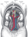
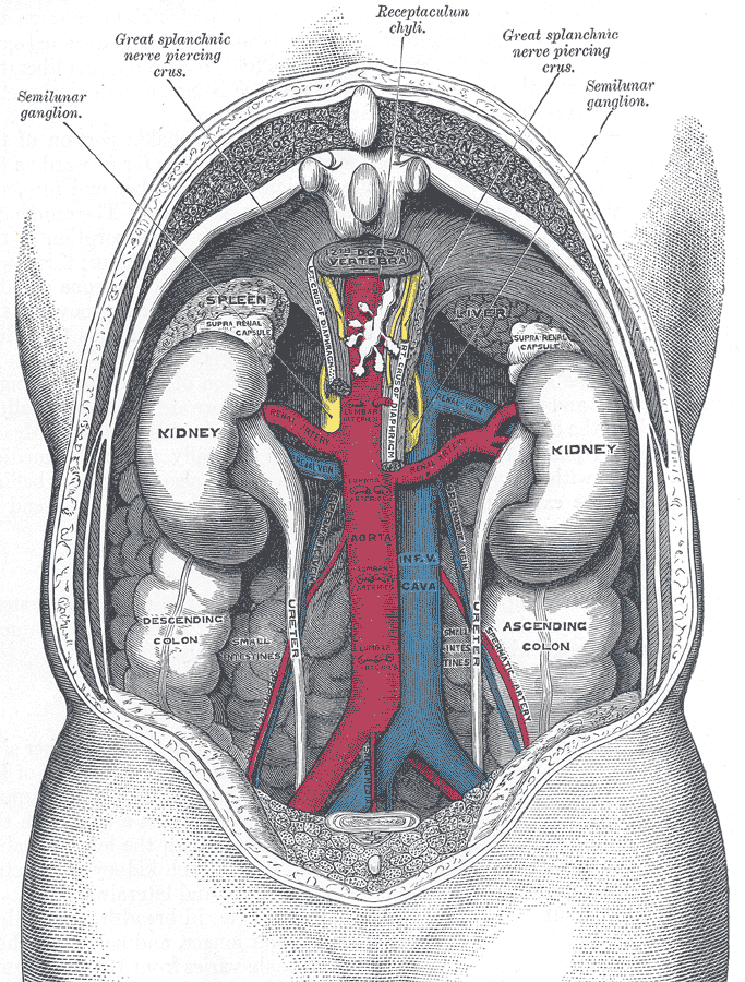
The relations of the viscera and large vessels of the abdomen. (Seen from behind, the last thoracic vertebra being well raised.)
External links
- SUNY Figs 40:04-16 - "The abdominal surface of the diaphragm."
- left+crus+of+diaphragm at eMedicine Dictionary
- right+crus+of+diaphragm at eMedicine Dictionary
This article was originally based on an entry from a public domain edition of Gray's Anatomy. As such, some of the information contained within it may be outdated.
Thoracic diaphragm (TA A04.4.02, GA 4.404) General Openings major: Caval opening · Esophageal hiatus · Aortic hiatus
minor: Sternocostal triangle · Lumbocostal triangle
This anatomy article is a stub. You can help Wikipedia by expanding it.