- Body of tibia
Infobox Bone
Name = Body of tibia
Latin = c. tibiae
GraySubject = 61
GrayPage = 256
Caption = Bones of the right leg. Anterior surface.

Caption2 = Bones of the right leg. Posterior surface.
System =
MeshName =
MeshNumber =
DorlandsPre = c_56
DorlandsSuf = 12260849
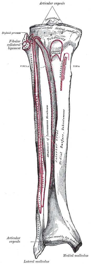
The body of thetibia has three borders and three surfaces.Borders
The anterior crest or border, the most prominent of the three, commences above at the tuberosity, and ends below at the anterior margin of the
medial malleolus . It is sinuous and prominent in the upper two-thirds of its extent, but smooth and rounded below; it gives attachment to the deepfascia of the leg.The medial border is smooth and rounded above and below, but more prominent in the center; it begins at the back part of the medial condyle, and ends at the posterior border of the medial malleolus; its upper part gives attachment to the tibial collateral ligament of the knee-joint to the extent of about 5 cm., and insertion to some fibers of the
Popliteus ; from its middle third some fibers of theSoleus andFlexor digitorum longus take origin.The interosseous crest or lateral border is thin and prominent, especially its central part, and gives attachment to the
interosseous membrane ; it commences above in front of the fibular articular facet, and bifurcates below, to form the boundaries of a triangular rough surface, for the attachment of the interosseous ligament connecting the tibia and fibula.Surfaces
The medial surface is smooth, convex, and broader above than below; its upper third, directed forward and medialward, is covered by the
aponeurosis derived from the tendon of the sartorius, and by the tendons of theGracilis andSemitendinosus , all of which are inserted nearly as far forward as the anterior crest; in the rest of its extent it issubcutaneous .The lateral surface is narrower than the medial; its upper two-thirds present a shallow groove for the origin of the Tibialis anterior; its lower third is smooth, convex, curves gradually forward to the anterior aspect of the bone, and is covered by the tendons of the
Tibialis anterior ,Extensor hallucis longus , andExtensor digitorum longus , arranged in this order from the medial side.The posterior surface presents, at its upper part, a prominent ridge, the popliteal line, which extends obliquely downward from the back part of the articular facet for the fibula to the medial border, at the junction of its upper and middle thirds; it marks the lower limit of the insertion of the
Popliteus , serves for the attachment of the fascia covering this muscle, and gives origin to part of theSoleus ,Flexor digitorum longus , andTibialis posterior . The triangular area, above this line, gives insertion to the Popliteus. The middle third of the posterior surface is divided by a vertical ridge into two parts; the ridge begins at the popliteal line and is well-marked above, but indistinct below; the medial and broader portion gives origin to the Flexor digitorum longus, the lateral and narrower to part of theTibialis posterior . The remaining part of the posterior surface is smooth and covered by the Tibialis posterior,Flexor digitorum longus , andFlexor hallucis longus . Immediately below the popliteal line is the nutrient foramen, which is large and directed obliquely downward.
Wikimedia Foundation. 2010.