- Superior olivary complex
-
For the cerebellar structure, see Dentate nucleus.
Brain: Superior olivary complex 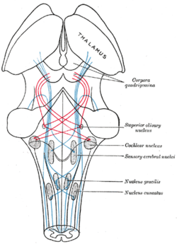
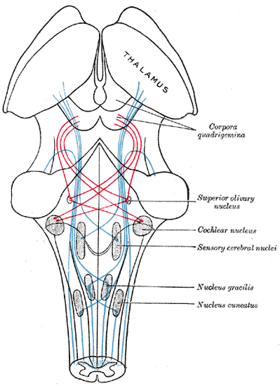
Scheme showing the course of the fibers of the lemniscus; medial lemniscus in blue, lateral in red. (Superior olivary nucleus is labeled at center right.) Latin nucleus olivaris superior Gray's subject #187 787 NeuroLex ID birnlex_1307 The superior olivary complex (or SOC or superior olive) is a collection of brainstem nuclei that functions in multiple aspects of hearing and is an important component of the ascending and descending auditory pathways. The SOC is intimately related to the trapezoid body: most of the cell groups of the SOC are dorsal (posterior in primates) to this axon bundle while a number of cell groups are embedded in the trapezoid body. Overall, the SOC displays a significant interspecies variation, being largest in bats and rodents and smaller in primates.
Physiology
The superior olivary nucleus plays a number of roles in hearing. The medial superior olive (MSO) is a specialized nucleus that is believed to measure the time difference of arrival of sounds between the ears (the interaural time difference or ITD). The ITD is a major cue for determining the azimuth of low-frequency sounds, i.e., localising them on the azimuthal plane – their degree to the left or the right.
The lateral superior olive (LSO) is believed to be involved in measuring the difference in sound intensity between the ears (the interaural level difference or ILD). The ILD is a second major cue in determining the azimuth of high-frequency sounds.
Relationship to auditory system
The superior olivary complex is generally located in the pons, but in human extends from the rostral medulla to the mid-pons[1] and receives projections predominantly from the anteroventral cochlear nucleus via the trapezoid body, although the posteroventral nucleus projects to the SOC via the intermediate acoustic stria. The SOC is the first major site of convergence of auditory information from the left and right ears. [2]
Primary Nuclei
The superior olivary complex is divided into three primary nuclei, the MSO, LSO, and the Medial Nucleus of the Trapezoid body, and several smaller periolivary nuclei.[3] These three nuclei are the most studied, and therefore best understood. Typically, they are regarded as forming the ascending azimuthal localization pathway.
Medial superior olive (MSO)
The medial superior olive is thought to help locate the azimuth of a sound, that is, the angle to the left or right where the sound source is located. One’s first instincts may be to think that this nucleus includes vertical information, but this is not the case; the information processed in the MSO is restricted to the horizontal plane. The fusiform cells of the Dorsal cochlear nucleus, which are thought to contribute to localization in elevation, bypass the SOC and project directly to the inferior colliculus. Only horizontal data is present, but it does come from two different ear sources, which aids in the localizing of sound on the azimuth axis.[4] The way in which the superior olive does this is by measuring the differences in time between two ear signals recording the same stimulus. Traveling around the head takes about 700 μs, and it is assumed that the medial superior olive is able to detect this. In fact, it is observed that people can detect interaural differences down to 10 μs.[4] The nucleus is tonotopically organized, but the azimuthal receptive field projection is “most likely a complex, nonlinear map.”[5]
The projections of the medial superior olive terminate densely in the ipsilateral central nucleus of the inferior colliculus. The majority of these axons are considered to be “round shaped” or type R. These R axons are mostly glutamatergic and contain round synaptic vesicles and form asymmetric synaptic junctions.[2]
- This is the largest of the nuclei and in human contains approximately 15,500 neurons [6].
- Each MSO receives low-frequency bilateral inputs from the right and left AVCNs.
- The output is to the ipsilateral lateral lemniscus and ultimately to the inferior colliculus.
- The MSO responds better to binaural stimuli.
- Its main function involves detection of differences in arrival time of sounds to the two ears which is part of localization process (ITD).
- The MSO is severely disrupted in the autistic brain [7].
Lateral superior olive (LSO)
This olive has similar functions to the medial superior olive, but employs intensity to localize the sound source.[8] The LSO receives excitatory, glutamatergic input from spherical bushy cells in the ipsilateral cochlear nucleus and inhibitory, glycinergic input from the medial nucleus of the trapezoid body (MNTB). The MNTB is driven by excitatory input from globular bushy cells in the contralateral cochlear nucleus. Thus, the LSO receives excitatory input from the ipsilateral ear and inhibitory input from the contralateral ear. This is the basis of ILD sensitivity. Projections from both cochlear nuclei are primarily high frequency, and these frequencies are subsequently represented by the majority of LSO neurons (>2/3 over 2-3kHz in cat). It should be noted that the LSO does in fact encode frequency across the animals audible range (not just "high" frequency. Additional inputs derive from the ipsilateral LNTB (glycinergic, see below), which provide inhibitory information from the ipsilateral cochlear nucleus[9]. Another possibly inhibitory input derives from ipsilateral AVCN non-spherical cells. These cells are either globular bushy or multipolar (stellate). Either of these two inputs could provide the basis for ipsilateral inhibition seen in response maps flanking the primary excitation, sharpening the unit's frequency tuning.[10][11]
The LSO projects bilaterally to the central nucleus of the inferior colliculus (ICC). Ipsilateral projections are primarily inhibitory (glycinergic), and the contralateral projections are excitatory. Additional projection targets include the Dorsal and Ventral nuclei of the Lateral Lemniscus (DNLL & VNLL). The GABAergic projections from the DNLL form a major source of GABA in the auditory brainstem, and project bilaterally to the ICC and to the contralateral DNLL. These converging excitatory and inhibitory connections may act to decrease the level dependence of ILD sensitivity in the ICC compared to the LSO.
Additional projections form the Lateral Olivocochlear Bundle (LOC), which innervates cochlear inner hair cells. These projections are thought to have a long time constant, and act to normalize the sound level detected by each ear in order to aid in sound localization[12]. Considerable species differences exist: LOC projection neurons are distributed within the LSO in rodents, and surround the LSO in predators (i.e. cat).
Medial Nucleus of Trapezoid Body (MNTB)
- The MNTB is composed of mainly neurons with round cell bodies which utilize glycine as a neurotransmitter.
- The size of the MNTB is reduced in primates [13], [14], [15]
- Each MNTB neuron receives a large "calyx" type ending, the Calyx of held arising from the globular bushy cells in the contralateral AVCN.
- There are two response types found: a ‘chopper type’ similar to spindle cells in the AVCN and a primary type which is similar to those of Bushy Cells in the AVCN.
Periolivary Nuclei
The SOC is composed of between six and nine periolivary nuclei, depending upon the researcher cited, typically named based upon their location with regard to the primary nuclei. These nuclei surround each of the primary nuclei, and contribute to both the ascending and descending auditory systems. These nuclei also form the source of the olivocochlear bundle, which innervates the cochlea.[16] In the guinea pig, ascending projections to the inferior colliculi are primarily ipsilateral (>80%), with the largest single source coming from the SPON. Also, ventral nuclei (RPO, VMPO, AVPO, & VNTB) are almost entirely ipsilateral, while the remaining nuclei project bilaterally.[17]
Name Cat Guinea Pig Rat Mouse LSO X X X X MSO X X X X MNTB X X X X LNTB X X "LVPO" X ALPO X X PVPO X X PPO X X "CPO" VLPO X DPO X X X DLPO X X VTB X X "MVPO" X AVPO X VMPO X X RPO X X SPN "DMPO" X X X Ventral Nucleus of Trapezoid Body (VNTB)
- The VNTB is a small nucleus located laterally to the MNTB, and ventral to the MSO.[18]
- Made up of a heterogeneous population of cells, this nucleus projects to many auditory nuclei, and forms the medial olivocochlear bundle (MOC) which innervates cochlear outer hair cells [19]. These cells contain electromotile fibers, and act as mechanical amplifiers/attenuators within the cochlea.
- The nucleus projects to both IC, with no cells projecting bilaterally.[20]
Lateral Nucleus of the Trapezoid Body (LNTB)
- Located ventral to the LSO[18]
- AVCN spherical bushy cells project collaterals bilaterally, and globular bushy cells project collaterals ipsilaterally to LNTB neurons.[21]
- Cells are immunoreactive for glycine[22], and are retrogradely labeled following injection of tritiated glycine into the LSO[9]
- The nucleus projects to both IC, with few cells projecting bilaterally[20], as well as the ipsilateral LSO[9].
- Large multipolar cells project to the cochlear nucleus, but not the IC, in both cat and guinea pig.[20][23]
- Inputs are often via end-bulbs of Held, producing very fast signal transduction.
Superior Periolivary Nucleus (SPON) (Dorsomedial Periolivary Nucleus (DMPO))
- Located directly dorsal to the MNTB[18]
- In rat, SPON is a homogeneous GABAergic nucleus. These tonotopically organized neurons receive excitatory inputs from octopus and multipolar cells in the contralateral ventral cochlear nucleus[24], a glycinergic (inhibitory) input from the ipsilateral MNTB, an unknown GABAergic (inhibitory) input, and project to the ipsilateral ICC.[25] Most neurons respond only at the offset of a stimulus, can phase lock to AM stimuli up to 200 Hz, and may form the basis for ICC duration selectivity.[26] Notably, SPON neurons do not receive descending inputs from the IC, and it does not project to the cochlea or cochlear nucleus as many periolivary nuclei do.[27]
- In guinea pig, round to oval multipolar cells project to both IC, with many cells projecting bilaterally. The more elongated cells that project to the cochlear nucleus to not project to the ICC. There appear to be to populations of cells, one that projects ipsilaterally, and one that projects bilaterally.[20]
- The majority of information had come from rodent SPON, due to the nucleus' prominent size in these species, with very few studies have been done in cat DMPO[28], none of which were extensive.
Dorsal Periolivary Nucleus (DPO)
- Located dorsal and medial to the LSO[18]
- Contains both EE (excited by both ears) and E0 (excited by the contralateral ear only) units.
- Neurons are tonotopically organized, and high frequency.
- May belong to a single nucleus along with the DLPO
- The nucleus projects to both IC, with no cells projecting bilaterally.[20]
Dorsolateral Periolivary Nucleus (DLPO)
- Located dorsal and lateral to the LSO[18]
- Contains both EE (excited by both ears) and E0 (excited by the contralateral ear only) units.
- Neurons are tonotopically organized, and low frequency.
- May belong to a single nucleus along with the DPO
- The nucleus projects to both IC, with few cells projecting bilaterally.[20]
Ventrolateral Periolivary Nucleus (VLPO)
- Located ventral to and within the ventral hillus of the LSO[18]
- Contains both EI (excited by contralateral and inhibited by ipsilateral ear) and E0 (excited by the contralateral ear only) units.
- Neurons are tonotopically organized, and high frequency.
Anterolateral Periolivary Nucleus (ALPO)
- The nucleus projects to both IC, with no cells projecting bilaterally.[20]
- Large multipolar cells project to the cochlear nucleus, but not the IC, in both cat and guinea pig.[20][23]
Ventromedial Periolivary Nucleus (VMPO)
- Located between the MSO and MNTB.[18]
- Sends projections to the ICC bilaterally.[20]
- The nucleus projects to both IC, with no cells projecting bilaterally.[20]
Rostral Periolivary Nucleus (RPO) (Anterior Periolivary Nucleus (APO))
Caudal Periolivary Nucleus (CPO) (Posterior Periolivary Nucleus (PPO))
- Located between the caudal pole of the MSO and the facial nucleus (7N)[18]
Posteroventral Periolivary Nucleus (PVPO)
- The nucleus projects to both IC, with no cells projecting bilaterally.[20]
Pathophysiology
An autopsy of a 21-year-old woman with autism, epilepsy and mental retardation found a near-complete absence of the superior olive.[30]
See also
References
- ^ Kulesza RJ, Cytoarchitecture of the human superior olivary complex: Medial and lateral superior olive. Hearing Research 225(2007) 80-90
- ^ a b Oliver DL, et al. Axonal projections from the lateral and medial superior olive to the inferior colliculus of the cat: a study using electron microscopic autoradiography. J Comp Neurol. 1995 Sep 11;360(1):17-32
- ^ Cajal, S. R. Y. and L. Azoulay (1909). Histologie du système nerveux de l'homme et des vertébrés. Paris, Maloine.
- ^ a b Kandel, et al. Principles of Neural Science. Fourth ed. pp 591-624. Copyright 2000, by McGraw-Hill Co.
- ^ Oliver, Douglas L. et al. Topography of Interaural Temporal Disparity Coding in Projections of Medial Superior Olive to Inferior Colliculus. The Journal of Neuroscience, August 13, 2003, 23(19):7438-7449
- ^ Kulesza, R. J., Jr. (2007). "Cytoarchitecture of the human superior olivary complex: medial and lateral superior olive." Hear Res 225(1-2): 80-90.
- ^ Kulesza et al., (2011) Malformation of the human superior olive in autistic spectrum disorders. Brain Res. 2011 Jan 7;1367:360-71.
- ^ Tsuchitani, C. and J. C. Boudreau (1967). "Encoding of stimulus frequency and intensity by cat superior olive S-segment cells." J Acoust Soc Am 42(4): 794-805.
- ^ a b c Glendenning, K. K., R. B. Masterton, et al. (1991). "Acoustic chiasm. III: Nature, distribution, and sources of afferents to the lateral superior olive in the cat." J Comp Neurol 310(3): 377-400.
- ^ Wu, S. H. and J. B. Kelly (1994). "Physiological evidence for ipsilateral inhibition in the lateral superior olive: synaptic responses in mouse brain slice." Hear Res 73(1): 57-64.
- ^ Brownell, W. E., P. B. Manis, et al. (1979). "Ipsilateral inhibitory responses in the cat lateral superior olive." Brain Res 177(1): 189-93.
- ^ Darrow, K. N., S. F. Maison, et al. (2006). "Cochlear efferent feedback balances interaural sensitivity." Nat Neurosci 9(12): 1474-6.
- ^ Bazwinsky et al., Characterization of the rhesus monkey superior olivary complex by calcium binding proteins and synaptophysin.J Anat. 2005 Dec;207(6):745-61.
- ^ Bazwinsky et al., Characterization of the human superior olivary complex by calcium binding proteins and neurofilament H (SMI-32).J Comp Neurol. 2003 Feb 10;456(3):292-303.
- ^ Kulesza, Cytoarchitecture of the human superior olivary complex: nuclei of the trapezoid body and posterior tier.Hear Res. 2008 Jul;241(1-2):52-63. Epub 2008 May 10.
- ^ Warr, W. B. and J. J. Guinan, Jr. (1979). "Efferent innervation of the organ of corti: two separate systems." Brain Res 173(1): 152-5.
- ^ a b Schofield, B. R. and N. B. Cant (1991). "Organization of the superior olivary complex in the guinea pig. I. Cytoarchitecture, cytochrome oxidase histochemistry, and dendritic morphology." J Comp Neurol 314(4): 645-70.
- ^ a b c d e f g h i Illing, R. B., K. S. Kraus, et al. (2000). "Plasticity of the superior olivary complex." Microsc Res Tech 51(4): 364-81.
- ^ Warr, W. B. and J. E. Beck (1996). "Multiple projections from the ventral nucleus of the trapezoid body in the rat." Hear Res 93(1-2): 83-101.
- ^ a b c d e f g h i j k Schofield, B. R. and N. B. Cant (1992). "Organization of the superior olivary complex in the guinea pig: II. Patterns of projection from the periolivary nuclei to the inferior colliculus." J Comp Neurol 317(4): 438-55.
- ^ Smith, P. H., P. X. Joris, et al. (1993). "Projections of physiologically characterized spherical bushy cell axons from the cochlear nucleus of the cat: evidence for delay lines to the medial superior olive." J Comp Neurol 331(2): 245-60.
- ^ Wenthold, R. J., D. Huie, et al. (1987). "Glycine immunoreactivity localized in the cochlear nucleus and superior olivary complex." Neuroscience 22(3): 897-912.
- ^ a b Adams, J. C. (1983). "Cytology of periolivary cells and the organization of their projections in the cat." J Comp Neurol 215(3): 275-89.
- ^ Friauf, E. and J. Ostwald (1988). "Divergent projections of physiologically characterized rat ventral cochlear nucleus neurons as shown by intra-axonal injection of horseradish peroxidase." Exp Brain Res 73(2): 263-84.
- ^ Kulesza, R. J., Jr. and A. S. Berrebi (2000). "Superior paraolivary nucleus of the rat is a GABAergic nucleus." J Assoc Res Otolaryngol 1(4): 255-69.
- ^ Kulesza, R. J., Jr., G. A. Spirou, et al. (2003). "Physiological response properties of neurons in the superior paraolivary nucleus of the rat." J Neurophysiol 89(4): 2299-312.
- ^ WHITE JS, WARR WB. The dual origins of the olivocochlear bundle in the albino rat. J. Comp. Neurol. 219:203–214, 1983.
- ^ Guinan, J. J., Jr., S. S. Guinan, et al. (1972). "Single auditory units in the superior olivary complex. I. Responses to sounds and classifications based on physiological properties." Int J Neurosci 4: 101-20.
- ^ Tsuchitani, C. (1977). "Functional organization of lateral cell groups of cat superior olivary complex." J Neurophysiol 40(2): 296-318.
- ^ Rodier PM, Ingram JL, Tisdale B, Nelson S, Romano J (1996). "Embryological origin for autism: developmental anomalies of the cranial nerve motor nuclei". J Comp Neurol 370 (2): 247–61. doi:10.1002/(SICI)1096-9861(19960624)370:2<247::AID-CNE8>3.0.CO;2-2. PMID 8808733.
External links
This article was originally based on an entry from a public domain edition of Gray's Anatomy. As such, some of the information contained within it may be outdated.
Human brain, rhombencephalon, metencephalon: pons (TA A14.1.05.101–604, GA 9.785) Dorsal/
(tegmentum)SurfaceWhite: Sensory/ascendingTrapezoid body/VIII · Trigeminal lemniscus (Dorsal trigeminal tract, Ventral trigeminal tract) · Medial lemniscus · Lateral lemniscus
MLF, III, IV and VI: Vestibulo-oculomotor fibers
Anterior trigeminothalamic tract · Central tegmental tractWhite: Motor/descendingICP (Vestibulocerebellar tract)
MLF, III, IV and VI: Vestibulospinal tract (Medial vestibulospinal tract, Lateral vestibulospinal tract)Other greyApneustic center · Pneumotaxic center (Medial parabrachial nucleus) · Lateral parabrachial nucleus · Superior olivary nucleus · Caerulean nucleusVentral/
(base)White: Motor/descendingSurfaceBasilar sulcusOther grey: Raphe/
reticularAuditory and vestibular pathways Auditory inner ear: Hair cells → Spiral ganglion → Cochlear nerve VIII →
pons: Cochlear nuclei (Anterior, Dorsal) → Trapezoid body → Superior olivary nuclei →
midbrain: Lateral lemniscus → Inferior colliculi →
thalamus: Medial geniculate nuclei →
cerebrum: Acoustic radiation → Primary auditory cortexVestibular inner ear: Vestibular nerve VIII →
pons: Vestibular nuclei (Medial vestibular nucleus, Lateral vestibular nucleus)
cerebellum: Flocculonodular lobe
spinal cord: Vestibulospinal tract (Medial vestibulospinal tract, Lateral vestibulospinal tract)
thalamus: Ventral posterolateral nucleus
Vestibulo-oculomotor fibersM: EAR
anat(e/p)/phys/devp
noco/cong, epon
proc, drug(S2)
Categories:
Wikimedia Foundation. 2010.