- Meckel's cartilage
-
Meckel's cartilage 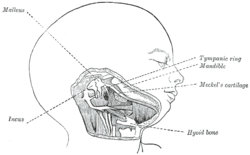
Head and neck of a human embryo eighteen weeks old, with Meckel’s cartilage and hyoid bar exposed. 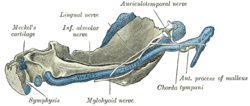
Mandible of human embryo 95 mm. long. Inner aspect. Nuclei of cartilage stippled. Latin cartilago arcus pharyngei primi Gray's subject #13 66 Precursor first branchial arch Gives rise to incus, malleus Code TE E4.0.3.3.3.1.3 The cartilaginous bar of the mandibular arch is formed by what are known as Meckel’s cartilages (right and left) also known as Meckelian cartilages; above this the incus and malleus are developed.
The dorsal end of each cartilage is connected with the ear-capsule and is ossified to form the malleus; the ventral ends meet each other in the region of the symphysis menti, and are usually regarded as undergoing ossification to form that portion of the mandible which contains the incisor teeth.
The intervening part of the cartilage disappears; the portion immediately adjacent to the malleus is replaced by fibrous membrane, which constitutes the sphenomandibular ligament, while from the connective tissue covering the remainder of the cartilage the greater part of the mandible is ossified.
Johann Friedrich Meckel, the Younger discovered this cartilage in 1820.
This article was originally based on an entry from a public domain edition of Gray's Anatomy. As such, some of the information contained within it may be outdated.
Contents
Additional images
-

Vertical section of the mandible of an early human fetus. X 25.
Evolution
The Meckelian Cartilage, also known as "Meckel's Cartilage", is a piece of cartilage from which the mandibles (lower jaws) of vertebrates evolved. Originally it was the lower of two cartilages which supported the first gill arch (nearest the front) in early fish. Then it grew longer and stronger, and acquired muscles capable of closing the developing jaw.[1]
In early fish and in chondrichthyans (cartilaginous fish such as sharks, which are not primitive in any sense of the word), the Meckelian Cartilage continued to be the main component of the lower jaw. But in the adult forms of osteichthyans (bony fish) and their descendants (amphibians, reptiles, birds, mammals), the cartilage was covered in bone - although in their embryos the jaw initially develops as the Meckelian Cartilage. In all tetrapods the cartilage partially ossifies (changes to bone) at the rear end of the jaw and becomes the articular bone, which forms part of the jaw joint in all tetrapods except mammals.[1]
External links
- palaeos.com
- synd/2049 at Who Named It?
References
Human development of head and neck (GA 1.65, TE E5.3, 5.4) Face Nasal placode · Nasal pit · nasal prominences (Lateral, Medial) · Intermaxillary segment
Frontonasal prominence · Maxillary prominence · Mandibular prominence (Meckel's cartilage)Oral cavity Primary palate · Secondary palateLateral lingual swelling · Tuberculum impar · Copula linguae · Hypopharyngeal eminence · Gustatory placodeGeneral
branchial apparatusCategories:- Skeletal system
- Musculoskeletal system stubs
- Developmental biology stubs
- Embryology
-


Wikimedia Foundation. 2010.