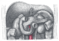
- Thoracic diaphragm
-
diaphragm 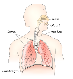
Respiratory system Latin diaphragma Artery Pericardiacophrenic artery, Musculophrenic artery, Inferior phrenic arteries Vein Superior phrenic vein, Inferior phrenic vein Nerve phrenic and lower intercostal nerves Precursor septum transversum, pleuroperitoneal folds, body wall [1] MeSH Diaphragm In the anatomy of mammals, the thoracic diaphragm, or simply the diaphragm (Ancient Greek: διάφραγμα diáphragma "partition"), is a sheet of internal skeletal muscle[2] that extends across the bottom of the rib cage. The diaphragm separates the thoracic cavity (heart, lungs & ribs) from the abdominal cavity and performs an important function in respiration. A diaphragm in anatomy can refer to other flat structures such as the urogenital diaphragm or pelvic diaphragm, but "the diaphragm" generally refers to the thoracic diaphragm. Other vertebrates such as amphibians and reptiles have diaphragms or diaphragm-like structures, but important details of the anatomy vary, such as the position of lungs in the abdominal cavity.
Contents
Function
The diaphragm functions in breathing. During inhalation, the diaphragm contracts, thus enlarging the thoracic cavity (the external intercostal muscles also participate in this enlargement). This reduces intra-thoracic pressure: In other words, enlarging the cavity creates suction that draws air into the lungs.
Cavity expansion happens in two extremes, along with intermediary forms. When the lower ribs are stabilized and the central tendon of the diaphragm is mobile, a contraction brings the insertion (central tendon) towards the origins and pushes the lower cavity towards the pelvis, allowing the thoracic cavity to expand downward. This is often called belly breathing. When the central tendon is stabilized and the lower ribs are mobile, a contraction lifts the origins (ribs) up towards the insertion (central tendon) which works in conjunction with other muscles to allow the ribs to slide and the thoracic cavity to expand laterally and upwards.
When the diaphragm relaxes, air is exhaled by elastic recoil of the lung and the tissues lining the thoracic cavity. Assisting this function with muscular effort (called forced exhalation) involves the internal intercostal muscles used in conjunction with the abdominal muscles, which act as an antagonist paired with the diaphragm's contraction.
The diaphragm is also involved in non-respiratory functions, helping to expel vomit, feces, and urine from the body by increasing intra-abdominal pressure, and preventing acid reflux by exerting pressure on the esophagus as it passes through the esophageal hiatus.
In some non-human animals, the diaphragm is not crucial for breathing; a cow, for instance, can survive fairly asymptomatically with diaphragmatic paralysis as long as no massive aerobic metabolic demands are made of it.
Anatomy
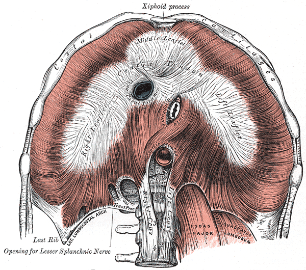
The diaphragm is a dome-shaped musculofibrous septum that separates the thoracic from the abdominal cavity, its convex upper surface forming the floor of the former, and its concave under surface forming the roof of the latter. Its peripheral part consists of muscular fibers that take origin from the circumference of the inferior thoracic aperture and converge to be inserted into a central tendon.
The muscular fibers may be grouped according to their origins into three parts:
Part Origin sternal two muscular slips from the back of the xiphoid process. costal the inner surfaces of the cartilages and adjacent portions of the lower six ribs on either side, interdigitating with the Transversus abdominis. lumbar aponeurotic arches, named the lumbocostal arches, and from the lumbar vertebrae by two pillars or crura. There are two lumbocostal arches, a medial and a lateral, on either side.
Innervation
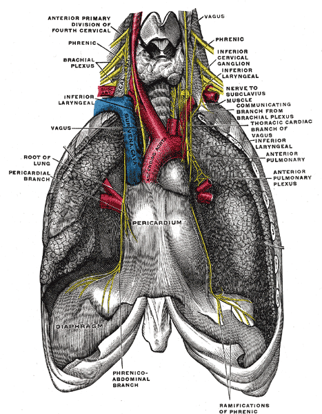
The diaphragm is primarily innervated by the phrenic nerve which is formed from the cervical nerves C3, C4, and C5. A useful mnemonic to remember this is, "C-3, 4, 5 keep the diaphragm alive." The peripheral portions of the diaphragm send sensory afferents via the intercostal (T5-T11) and subcostal nerves (T12).
Crura and central tendon
At their origins the crura are tendinous in structure, and blend with the anterior longitudinal ligament of the vertebral column.
The central tendon of the diaphragm is a thin but strong aponeurosis situated near the center of the vault formed by the muscle, but somewhat closer to the front than to the back of the thorax, so that the posterior muscular fibers are the longer.
Openings
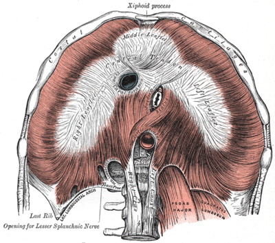 anterior view of the human diaphragm, showing openings
The diaphragm is pierced by a series of apertures to permit the passage of structures between the thorax and abdomen. Three large openings — the aortic, the esophageal, and the vena cava — and a series of smaller ones are described.
opening level structures caval opening T8 inferior vena cava, and some branches of the right phrenic nerve esophageal hiatus T10 esophagus, the anterior and posterior vagal trunks, and some small esophageal arteries aortic hiatus T12 the aorta, the azygos vein, and the thoracic duct two lesser aperture of right crus greater and lesser right splanchnic nerves three lesser aperture of left crus greater and lesser left splanchnic nerves and the hemiazygos vein behind the diaphragm, under the medial lumbocostal arches sympathetic trunk areolar tissue between the sternal and costal parts (see also foramina of Morgagni) the superior epigastric branch of the internal mammary artery and some lymphatics from the abdominal wall and convex surface of the liver areolar tissue between the fibers springing from the medial and lateral lumbocostal arches This interval is less constant; when this interval exists, the upper and back part of the kidney is separated from the pleura by areolar tissue only. 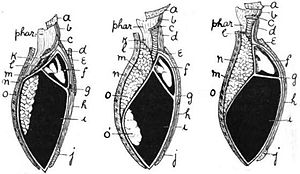 Diaphragm and pleural cavities in amphibian (left), bird (center), mammal (right). a, mandible; b, genio-hyoid; c, hyoid; d, sterno-hyoid; e, sternum; f, pericardium; g, septum transversum; h, rectus abdominis; i, abdominal cavity; j, pubis; k, esophagus; l, trachea; m, cervical limiting membrane of abdominal cavity; n, dorsal wall of body; o, lung; o', air-sac.[3]
A commonly used mnemonic to remember the level of the diaphragmatic apertures is this: Mnemonic
- Aortic hiatus = 12 letters = T12
- Oesophagus = 10 letters = T10
- Vena cava = 8 letters = T8
Another common mnemonic is: "I ate ten eggs at twelve" I (IVC) ate (TV8); ten (TV10) eggs (esophagus); at (aorta, azygos) twelve (TV12)
Comparative anatomy and evolution
The existence of some membrane separating the pharynx from the stomach can be traced widely among the chordates. Thus amphioxus possesses an atrium by which water exits the pharynx, which has been argued (and disputed) to be homologous to structures in ascidians and hagfishes.[4] The urochordate epicardium separates digestive organs from the pharynx and heart, but the anus returns to the upper compartment to discharge wastes through an outgoing siphon.
Thus the diaphragm emerges in the context of a body plan that separated an upper feeding compartment from a lower digestive tract, but the point at which it originates is a matter of definition. Structures in fish, amphibians, reptiles, and birds have been called diaphragms, but it has been argued that these structures are not homologous. For instance, the alligator diaphragmaticus muscle does not insert on the esophagus and does not affect pressure of the lower esophageal sphincter.[5] The lungs are located in the abdominal compartment of amphibians and reptiles, so that contraction of the diaphragm expels air from the lungs rather than drawing it into them. In birds and mammals, lungs are located above the diaphragm. The presence of an exceptionally well-preserved fossil of Sinosauropteryx, with lungs located beneath the diaphragm as in crocodiles, has been used to argue that dinosaurs could not have sustained an active warm-blooded physiology, or that birds could not have evolved from dinosaurs.[6] An explanation for this state of affairs is that lungs originated beneath the diaphragm, but as the demands for respiration increased in warm-blooded birds and mammals, natural selection came to favor the parallel evolution of the herniation of the lungs from the abdominal cavity in both lineages.[3] However, birds do not have diaphragms. They do not breathe in the same way as mammals, and do not rely on creating a negative pressure in the thoracic cavity, at least not to the same extent. They rely on a rocking motion of the keel of the sternum to create local areas of reduced pressure to supply thin, membranous airsacs cranially and caudally to the fixed-volume, non-expansive lungs. A complicated system of valves and air sacs cycles air constantly over the absorption surfaces of the lungs so allowing maximal efficiency of gaseous exchange. Thus, birds do not have the reciprocal tidal breathing flow of mammals. On careful dissection, around eight air sacs can be clearly seen. They extend quite far caudally into the abdomen.[7]
Variations
The sternal portion of the muscle is sometimes wanting and more rarely defects occur in the lateral part of the central tendon or adjoining muscle fibers.
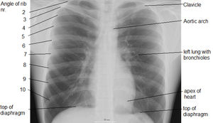 X-ray of chest, showing top of diaphragm.
Pathology
The right crus of the diaphragm is part of the lower esophageal sphincter (LES), which separates the thoracic and abdominal parts of the esophagus. A hiatal hernia, in which the abdominal esophagus or even the fundus of the stomach rises up through the esophageal hiatus into the thoracic cavity, can result from a tear or weakness in the diaphragm. Weakness may also result from injury to the neck at levels C3, C4, C5 which can result in altered tone of either half of the diaphragm. Both general weakness of the LES and hiatal hernia can cause "acid reflux," also known as gastroesophageal reflux disease u(GERD).
If the diaphragm is struck, or otherwise spasms, breathing will become difficult. This is called "having the wind knocked out of you."
Hiccoughs are involuntary intermittent sudden contractions of the diaphragm.
Gas inferior to the diaphragm in a human (or posterior in a four-footed creature) may be pneumoperitoneum, which can be a sign of serious pathology.
Development
The thoracic diaphragm develops embryologically beginning in the third week after fertilization with two processes known as transverse folding and longitudinal folding. The septum transversum, the primitive central tendon of the diaphragm, originates at the rostral pole of the embryo and is relocated during longitudinal folding to the ventral thoracic region. Transverse folding brings the body wall anteriorly to enclose the gut and body cavities. The pleuroperitoneal membrane and body wall myoblasts, from somatic lateral plate mesoderm, meet the septum transversum to close off the pericardio-peritoneal canals on either side of the presumptive esophagus, forming a barrier that separates the peritoneal and pleuropericardial cavities. Furthermore, dorsal mesenchyme surrounding the presumptive esophagus form the muscular crura of the diaphragm.
Because the earliest element of the embryological diaphragm, the septum transversum, forms in the cervical region, the phrenic nerve that innervates the diaphragm originates from the cervical spinal cord (C3,4, and 5). As the septum transversum descends inferiorly, the phrenic nerve follows, accounting for its circuitous route from the upper cervical vertebrae, around the pericardium, finally to innervate the diaphragm.
Clinical Relevance
Failure of one of the pleuroperitoneal membrane to form of a competent barrier between cavities can result in congenital diaphragmatic hernia. This condition is present in 1 out of 2,000 births. A large herniation has a mortality rate of 3/4 and requires immediate surgical repair.
Additional images
-
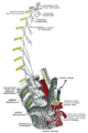
The phrenic nerve and its relations with the vagus nerve.
-
Plan of right sympathetic cord and splanchnic nerves.
-

Thoracic portion of the sympathetic trunk.
-
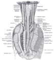
The position and relation of the esophagus in the cervical region and in the posterior mediastinum. Seen from behind.
-
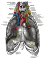
Front view of the thoracic and abdominal viscera.
-
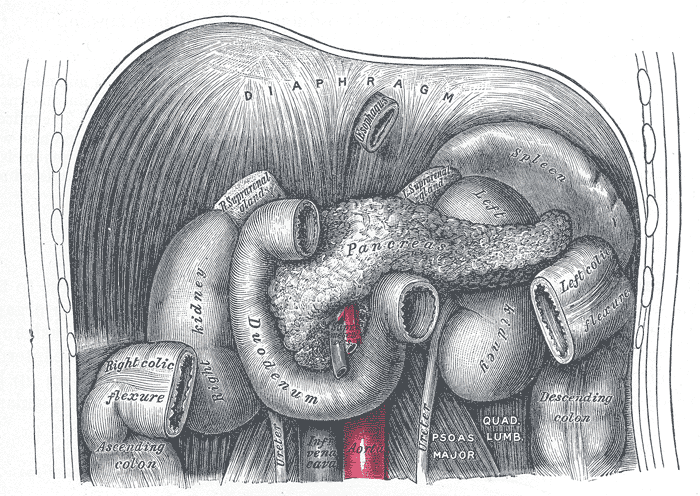
The duodenum and pancreas.
-
Topography of thoracic and abdominal viscera.
-

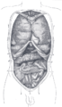
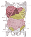
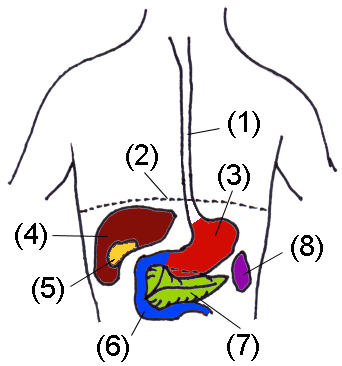
Organs of abdomen
-
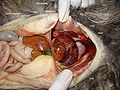
Peritoneopericardial diaphragmatic hernia in a cat.
-
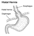
Hiatal hernia
See also
References
- ^ mslimb-012 — Embryology at UNC
- ^ Campbell, Neil A. (2009). Biology: Australian Version (8th ed.). Sydney: Pearson/Benjamin Cummings. pp. 334. ISBN 978-1-4425-0221-5.
- ^ a b Arthur Keith, M.D. (1905). The nature of the mammalian diaphragm and pleural cavities. http://books.google.com/books?id=2ecDAAAAYAAJ.
- ^ Zbynek Kozmik et al. (1999). "Characterization of an amphioxus paired box gene, AmphiPax2/5/8". Development 126 (6): 1295–1304. PMID 10021347. http://dev.biologists.org/cgi/reprint/126/6/1295.pdf.
- ^ T. J. Uriona et al. (2005). "Structure and function of the esophagus of the American alligator (Alligator mississippiensis)". Journal of Experimental Biology 208 (Pt 16): 3047–3053. doi:10.1242/jeb.01746. PMID 16081603. http://jeb.biologists.org/cgi/content/full/208/16/3047.
- ^ "Lung fossils suggest that dinos breathed in cold blood". http://cas.bellarmine.edu/tietjen/images/lung_fossils_suggest_dinos_breat.htm.
- ^ Dyce, Sack and Wensing in Textbook of Veterinary Anatomy; 2002 (3rd Edn); Saunders, Philiadelphia
 This article incorporates content from the 1728 Cyclopaedia, a publication in the public domain.
External links
This article was originally based on an entry from a public domain edition of Gray's Anatomy. As such, some of the information contained within it may be outdated.
Thoracic diaphragm (TA A04.4.02, GA 4.404) General Openings major: Caval opening · Esophageal hiatus · Aortic hiatus
minor: Sternocostal triangle · Lumbocostal triangleHuman systems and organs TA 2–4:
MSBone (Carpus · Collar bone (clavicle) · Thigh bone (femur) · Fibula · Humerus · Mandible · Metacarpus · Metatarsus · Ossicles · Patella · Phalanges · Radius · Skull (cranium) · Tarsus · Tibia · Ulna · Rib · Vertebra · Pelvis · Sternum) · CartilageTA 5–11:
splanchnic/
viscusmostly
Thoracicmostly
AbdominopelvicDigestive system+
adnexaMouth (Salivary gland, Tongue) · upper GI (Oropharynx, Laryngopharynx, Esophagus, Stomach) · lower GI (Small intestine, Appendix, Colon, Rectum, Anus) · accessory (Liver, Biliary tract, Pancreas)TA 12–16 Blood
(Non-TA)General anatomy: systems and organs, regional anatomy, planes and lines, superficial axial anatomy, superficial anatomy of limbsCategories:- Thorax
- Muscles of the torso


Wikimedia Foundation. 2010.