- Neuroimaging
-
 Para-sagittal MRI of the head in a patient with benign familial macrocephaly.
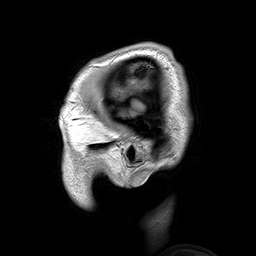
 3-D MRI of a section of the head.
Neuroimaging includes the use of various techniques to either directly or indirectly image the structure, function/pharmacology of the brain. It is a relatively new discipline within medicine and neuroscience/psychology.[1]
Contents
Overview
Neuroimaging falls into two broad categories:
- Structural imaging, which deals with the structure of the brain and the diagnosis of gross (large scale) intracranial disease (such as tumor), and injury, and
- functional imaging, which is used to diagnose metabolic diseases and lesions on a finer scale (such as Alzheimer's disease) and also for neurological and cognitive psychology research and building brain-computer interfaces.
Functional imaging enables, for example, the processing of information by centers in the brain to be visualized directly. Such processing causes the involved area of the brain to increase metabolism and "light up" on the scan. One of the more controversial uses of neuroimaging has been research into "Thought identification" or mind-reading.
History
Main article: History of neuroimagingIn 1918 the American neurosurgeon Walter Dandy introduced the technique of ventriculography. X-ray images of the ventricular system within the brain were obtained by injection of filtered air directly into one or both lateral ventricles of the brain. Dandy also observed that air introduced into the subarachnoid space via lumbar spinal puncture could enter the cerebral ventricles and also demonstrate the cerebrospinal fluid compartments around the base of the brain and over its surface. This technique was called pneumoencephalography.
In 1927 Egas Moniz introduced cerebral angiography, whereby both normal and abnormal blood vessels in and around the brain could be visualized with great precision.
In the early 1970s, Allan McLeod Cormack and Godfrey Newbold Hounsfield introduced computerized axial tomography (CAT or CT scanning), and ever more detailed anatomic images of the brain became available for diagnostic and research purposes. Cormack and Hounsfield won the 1979 Nobel Prize for Physiology or Medicine for their work. Soon after the introduction of CAT in the early 1980s, the development of radioligands allowed single photon emission computed tomography (SPECT) and positron emission tomography (PET) of the brain.
More or less concurrently, magnetic resonance imaging (MRI or MR scanning) was developed by researchers including Peter Mansfield and Paul Lauterbur, who were awarded the Nobel Prize for Physiology or Medicine in 2003. In the early 1980s MRI was introduced clinically, and during the 1980s a veritable explosion of technical refinements and diagnostic MR applications took place. Scientists soon learned that the large blood flow changes measured by PET could also be imaged by the correct type of MRI. Functional magnetic resonance imaging (fMRI) was born, and since the 1990s, fMRI has come to dominate the brain mapping field due to its low invasiveness, lack of radiation exposure, and relatively wide availability. As noted above fMRI is also beginning to dominate the field of stroke treatment.
In early 2000s the field of neuroimaging reached the stage where limited practical applications of functional brain imaging have become feasible. The main application area is crude forms of brain-computer interface.
Brain imaging techniques
Computed axial tomography
Main article: CT headComputed tomography (CT) or Computed Axial Tomography (CAT) scanning uses a series of x-rays of the head taken from many different directions. Typically used for quickly viewing brain injuries, CT scanning uses a computer program that performs a numerical integral calculation (the inverse Radon transform) on the measured x-ray series to estimate how much of an x-ray beam is absorbed in a small volume of the brain. Typically the information is presented as cross sections of the brain.[2]
Diffuse optical imaging
Diffuse optical imaging (DOI) or diffuse optical tomography (DOT) is a medical imaging modality which uses near infrared light to generate images of the body. The technique measures the optical absorption of haemoglobin, and relies on the absorption spectrum of haemoglobin varying with its oxygenation status.
Event-related optical signal (EROS) is a brain-scanning technique which uses infrared light through optical fibers to measure changes in optical properties of active areas of the cerebral cortex. Whereas techniques such as diffuse optical imaging (DOT) and near infrared spectroscopy (NIRS) measure optical absorption of haemoglobin, and thus are based on blood flow, EROS takes advantage of the scattering properties of the neurons themselves, and thus provides a much more direct measure of cellular activity. EROS can pinpoint activity in the brain within millimeters (spatially) and within milliseconds (temporally). Its biggest downside is the inability to detect activity more than a few centimeters deep. EROS is a new, relatively inexpensive technique that is non-invasive to the test subject. It was developed at the University of Illinois at Urbana-Champaign where it is now used in the Cognitive Neuroimaging Laboratory of Dr. Gabriele Gratton and Dr. Monica Fabiani.
Magnetic resonance imaging
Main article: MRI of brain and brain stem Sagittal MRI slice at the midline.
Magnetic resonance imaging (MRI) uses magnetic fields and radio waves to produce high quality two- or three-dimensional images of brain structures without use of ionizing radiation (X-rays) or radioactive tracers.
Functional magnetic resonance imaging
 Axial MRI slice at the level of the basal ganglia, showing fMRI BOLD signal changes overlayed in red (increase) and blue (decrease) tones.
Functional magnetic resonance imaging (fMRI) relies on the paramagnetic properties of oxygenated and deoxygenated hemoglobin to see images of changing blood flow in the brain associated with neural activity. This allows images to be generated that reflect which brain structures are activated (and how) during performance of different tasks.
Most fMRI scanners allow subjects to be presented with different visual images, sounds and touch stimuli, and to make different actions such as pressing a button or moving a joystick. Consequently, fMRI can be used to reveal brain structures and processes associated with perception, thought and action. The resolution of fMRI is about 2-3 millimeters at present, limited by the spatial spread of the hemodynamic response to neural activity. It has largely superseded PET for the study of brain activation patterns. PET, however, retains the significant advantage of being able to identify specific brain receptors (or transporters) associated with particular neurotransmitters through its ability to image radiolabelled receptor "ligands" (receptor ligands are any chemicals that stick to receptors).
As well as research on healthy subjects, fMRI is increasingly used for the medical diagnosis of disease. Because fMRI is exquisitely sensitive to blood flow, it is extremely sensitive to early changes in the brain resulting from ischemia (abnormally low blood flow), such as the changes which follow stroke. Early diagnosis of certain types of stroke is increasingly important in neurology, since substances which dissolve blood clots may be used in the first few hours after certain types of stroke occur, but are dangerous to use afterwards. Brain changes seen on fMRI may help to make the decision to treat with these agents. With between 72% and 90% accuracy where chance would achieve 0.8%,[3] fMRI techniques can decide which of a set of known images the subject is viewing.[4]
Magnetoencephalography
Magnetoencephalography (MEG) is an imaging technique used to measure the magnetic fields produced by electrical activity in the brain via extremely sensitive devices such as superconducting quantum interference devices (SQUIDs). MEG offers a very direct measurement of neural electrical activity (compared to fMRI for example) with very high temporal resolution but relatively low spatial resolution. The advantage of measuring the magnetic fields produced by neural activity is that they are likely to be less distorted by surrounding tissue (particularly the skull and scalp) compared to the electric fields measured by EEG. Specifically, it can be shown that magnetic fields produced by electrical activity are not affected by the surrounding head tissue, when the the head is modeled as a set of concentric spherical shells, each being an isotropic homogeneous conductor. Real heads are non-spherical and have largely anisotropic conductivities (particularly white matter and skull). While skull anisotropy has negligible effect on MEG (unlike EEG), white matter anisotropy strongly affects MEG measurements for radial and deep sources[5]. Note, however, that the skull was assumed to be uniformly anisotropic in this study, which is not true for a real head: the absolute and relative thicknesses of diploë and tables layers vary among and within the skull bones. This makes it likely that MEG is also affected by the skull anisotropy, although probably not to the same degree as EEG.
There are many uses for the MEG, including assisting surgeons in localizing a pathology, assisting researchers in determining the function of various parts of the brain, neurofeedback, and others.
Positron emission tomography
 PET scan of a normal 20-year-old brain.
Positron emission tomography (PET) measures emissions from radioactively labeled metabolically active chemicals that have been injected into the bloodstream. The emission data are computer-processed to produce 2- or 3-dimensional images of the distribution of the chemicals throughout the brain. [6] The positron emitting radioisotopes used are produced by a cyclotron, and chemicals are labeled with these radioactive atoms. The labeled compound, called a radiotracer, is injected into the bloodstream and eventually makes its way to the brain. Sensors in the PET scanner detect the radioactivity as the compound accumulates in various regions of the brain. A computer uses the data gathered by the sensors to create multicolored 2- or 3-dimensional images that show where the compound acts in the brain. Especially useful are a wide array of ligands used to map different aspects of neurotransmitter activity, with by far the most commonly used PET tracer being a labeled form of glucose (see Fludeoxyglucose (18F) (FDG)).
The greatest benefit of PET scanning is that different compounds can show blood flow and oxygen and glucose metabolism in the tissues of the working brain. These measurements reflect the amount of brain activity in the various regions of the brain and allow to learn more about how the brain works. PET scans were superior to all other metabolic imaging methods in terms of resolution and speed of completion (as little as 30 seconds), when they first became available. The improved resolution permitted better study to be made as to the area of the brain activated by a particular task. The biggest drawback of PET scanning is that because the radioactivity decays rapidly, it is limited to monitoring short tasks. [7] Before fMRI technology came online, PET scanning was the preferred method of functional (as opposed to structural) brain imaging, and it still continues to make large contributions to neuroscience.
PET scanning is also used for diagnosis of brain disease, most notably because brain tumors, strokes, and neuron-damaging diseases which cause dementia (such as Alzheimer's disease) all cause great changes in brain metabolism, which in turn causes easily detectable changes in PET scans. PET is probably most useful in early cases of certain dementias (with classic examples being Alzheimer's disease and Pick's disease) where the early damage is too diffuse and makes too little difference in brain volume and gross structure to change CT and standard MRI images enough to be able to reliably differentiate it from the "normal" range of cortical atrophy which occurs with aging (in many but not all) persons, and which does not cause clinical dementia.
Single photon emission computed tomography
Single photon emission computed tomography (SPECT) is similar to PET and uses gamma ray emitting radioisotopes and a gamma camera to record data that a computer uses to construct two- or three-dimensional images of active brain regions[8] SPECT relies on an injection of radioactive tracer, which is rapidly taken up by the brain but does not redistribute. Uptake of SPECT agent is nearly 100% complete within 30 – 60s, reflecting cerebral blood flow (CBF) at the time of injection. These properties of SPECT make it particularly well suited for epilepsy imaging, which is usually made difficult by problems with patient movement and variable seizure types. SPECT provides a "snapshot" of cerebral blood flow since scans can be acquired after seizure termination (so long as the radioactive tracer was injected at the time of the seizure). A significant limitation of SPECT is its poor resolution (about 1 cm) compared to that of MRI.
Like PET, SPECT also can be used to differentiate different kinds of disease processes which produce dementia, and it is increasingly used for this purpose. Neuro-PET has a disadvantage of requiring use of tracers with half-lives of at most 110 minutes, such as FDG. These must be made in a cyclotron, and are expensive or even unavailable if necessary transport times are prolonged more than a few half-lives. SPECT, however, is able to make use of tracers with much longer half-lives, such as technetium-99m, and as a result, is far more widely available.
See also
- Brain mapping
- Functional neuroimaging
- functional near-infrared imaging
- History of brain imaging
- Human Cognome Project
- Magnetic resonance imaging
- Magnetoencephalography
- Medical imaging
- List of neuroscience databases
- Neuroimaging software
- Statistical parametric mapping
- Transcranial magnetic stimulation
- Voxel-based morphometry
References
- ^ Filler, AG: The history, development, and impact of computed imaging in neurological diagnosis and neurosurgery: CT, MRI, DTI: Nature Precedings DOI: 10.1038/npre.2009.3267.5.Neurosurgical Focus (in press)
- ^ Malcom Jeeves (1994). Mind Fields: Reflections on the Science of Mind and Brain. Grand Rapids, MI: Baker Books., p. 21
- ^ Smith, Kerri (March 5, 2008). "Mind-reading with a brain scan". Nature News (Nature Publishing Group). http://www.nature.com/news/2008/080305/full/news.2008.650.html. Retrieved 2008-03-05.
- ^ Keim, Brandon (March 5, 2008). "Brain Scanner Can Tell What You're Looking At". Wired News (CondéNet). http://www.wired.com/science/discoveries/news/2008/03/mri_vision. Retrieved 2008-03-05.
- ^ Wolters et al, "Influence of tissue conductivity anisotropy on EEG/MEG field and return current computation in a realistic head model: A simulation and visualization study using high-resolution finite element modeling, NeuroImage, 30(3):813-26 (2006)"
- ^ Lars-Goran Nilsson and Hans J. Markowitsch (1999). Cognitive Neuroscience of Memory. Seattle: Hogrefe & Huber Publishers., page 57
- ^ Lars-Goran Nilsson and Hans J. Markowitsch (1999). Cognitive Neuroscience of Memory. Seattle: Hogrefe & Huber Publishers., pg. 60
- ^ Philip Ball Brain Imaging Explained
External links
- The Whole Brain Atlas @ Harvard
- The McConnell Brain Imaging Center, McGill University
- The American Society of Neuroimaging (ASN).
- UCLA Neuroimaging Training Program.
- Laboratory of Neuro Imaging at UCLA
- A Neuroimaging portal
- BrainMapping.org, a free BrainMapping community information portal
- Lecture notes on mathematical aspects of neuroimaging by Will Penny, University College London
- "Transcranial Magnetic Stimulation". by Michael Leventon in association with MIT AI Lab.
- Foundations of fMRI by Jamie Shorey.
- International Society for Neuroimaging in Psychiatry (ISNIP)
- Journal of Neuroimaging
Psychiatry Portal Subspecialties Addiction psychiatry · Biological psychiatry · Child and adolescent psychiatry · Cross-cultural psychiatry · Developmental disability · Eating disorders · Emergency psychiatry · Forensic psychiatry · Geriatric psychiatry · Liaison psychiatry · Military psychiatry · Neuropsychiatry · Palliative medicine · Pain medicine · Psychotherapy · Sleep medicine
Organizations American Board of Psychiatry and Neurology · American Psychiatric Association · American Neuropsychiatric Association · Brazilian Association of Psychiatry · Canadian Psychiatric Association · Chinese Society of Psychiatry · Democratic Psychiatry · German Society of Psychiatry, Psychotherapy and Neurology · Hong Kong College of Psychiatrists · Independent Psychiatric Association of Russia · Indian Psychiatric Society · Irish College of Psychiatrists · Israeli Psychiatric Association · Italian Psychiatric Society · Japanese Society of Psychiatry & Neurology · Korean Neuropsychiatric Association · Maryland Psychiatric Society · National Institute of Mental Health · Pakistan Psychiatric Society · Royal Australian and New Zealand College of Psychiatrists · Royal College of Psychiatrists · Singapore Psychiatric Association · South African Society of Psychiatrists · World Psychiatric Association
Related topics Anti-psychiatry · Behavioral medicine · Clinical neuroscience · Imaging genetics · Neuroimaging · Neurophysiology · Psychiatrist · Psychiatric epidemiology · Psychiatric genetics · Psychiatric survivors movement · Psychosomatic medicine · Psycho-oncology · Psychopharmacology · Psychosurgery · Psychoanalysis
Lists Visualization of technical information Fields Biological data visualization · Chemical imaging · Crime mapping · Data visualization · Educational visualization · Flow visualization · Geovisualization · Information visualization · Mathematical visualization · Medical imaging · Molecular graphics · Product visualization · Scientific visualization · Software visualization · Technical drawing · Visual culture · Volume visualizationImage types Experts Jacques Bertin · Stuart Card · Thomas A. DeFanti · Michael Friendly · Nigel Holmes · Alan MacEachren · Jock D. Mackinlay · Michael Maltz · Bruce H. McCormick · Charles Joseph Minard · Otto Neurath · William Playfair · Clifford A. Pickover · Arthur H. Robinson · Lawrence J. Rosenblum · Adolphe Quetelet · George G. Robertson · Ben Shneiderman · Edward TufteRelated topics Cartography · Computer graphics · Computer graphics (computer science) · Model-based definition · Graph drawing · Graphic design · Graphic organizer · Imaging science · Information graphics · Information science · Mental visualisation · Neuroimaging · Scientific modelling · Spatial analysis · Visual analytics · Visual perceptionSurgery, Nervous system: neurosurgical and other procedures (ICD-9-CM V3 01–05+89.1, ICD-10-PCS 00-01) Skull CNS thalamus and globus pallidus: Thalamotomy · Thalamic stimulator · Pallidotomy
ventricular system: Ventriculostomy · Suboccipital puncture · Intracranial pressure monitoring
cerebrum: Psychosurgery (Lobotomy, Bilateral cingulotomy) · Hemispherectomy · Anterior temporal lobectomy
pituitary: Hypophysectomy
hippocampus: Amygdalohippocampectomy
Brain biopsyCerebral meningesSpinal cord and roots (Cordotomy, Rhizotomy)
Vertebrae and intervertebral discs: see Template:Bone, cartilage, and joint proceduresCT head · Cerebral angiography · Pneumoencephalography · Echoencephalography/Transcranial doppler · MRI of brain and brain stem · Brain PET · SPECT of brain · MyelographyDiagnosticPNS Sympathetic nerves or gangliaNerves (general)DiagnosticCategories:


Wikimedia Foundation. 2010.