- Papanicolaou stain
-
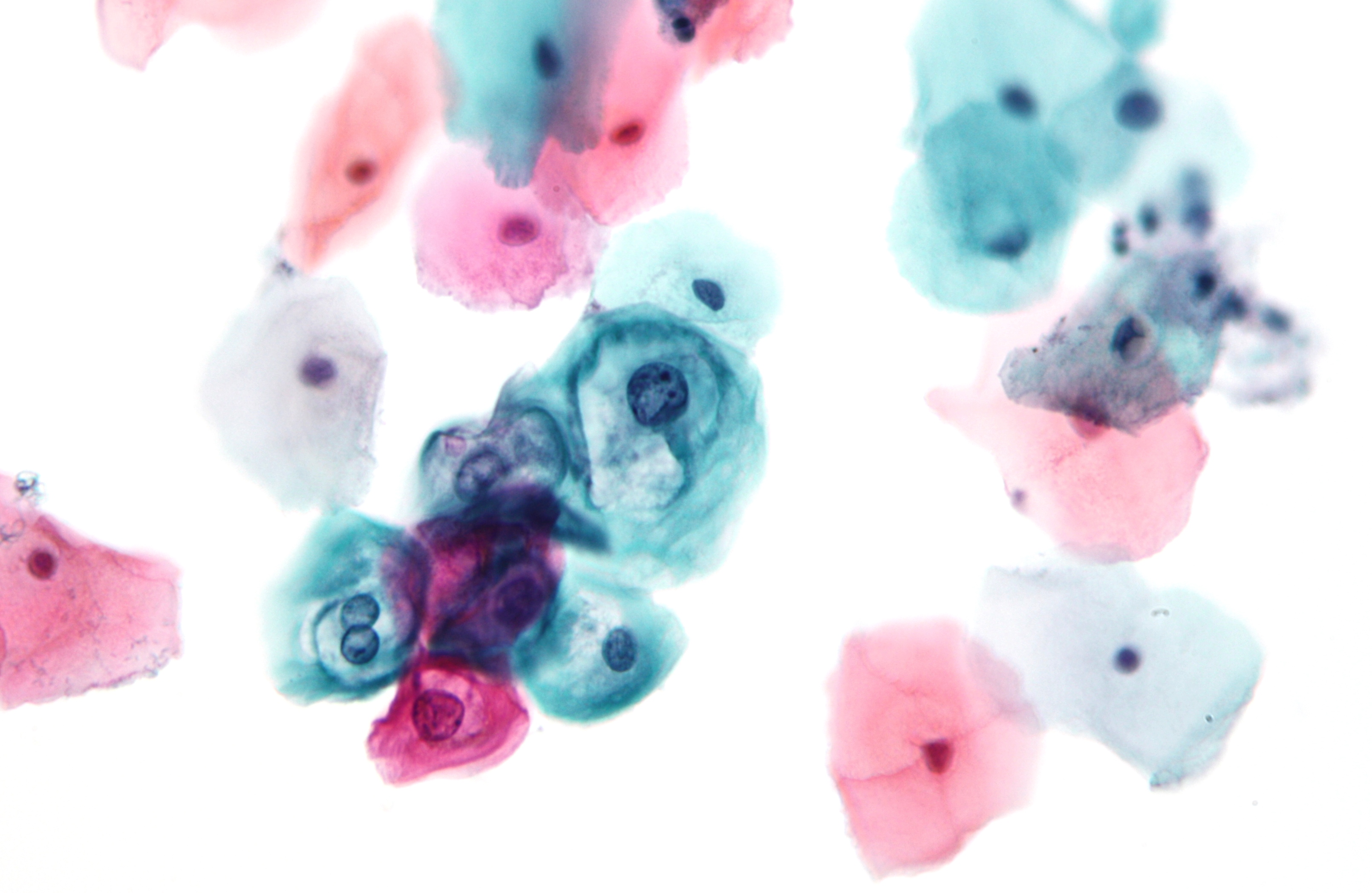
 Papanicolaou stain showing a low-grade squamous intraepithelial lesion (LSIL). Pap test.
Papanicolaou stain (also Papanicolaou's stain and Pap stain) is a multichromatic staining histological technique developed by George Papanikolaou, the father of cytopathology.
Pap staining is used to differentiate cells in smear preparations of various bodily secretions; the specimens can be gynecological smears (Pap smears), sputum, brushings, washings, urine, cerebrospinal fluid, abdominal fluid, pleural fluid, synovial fluid, seminal fluid, fine needle aspiration material, tumor touch samples, or other materials containing cells.
Pap staining is a very reliable technique. As such, it is used for cervical cancer screening in gynecology. The entire procedure is known as Pap smear.
The classic form of Pap stain involves five dyes in three solutions:[1]
- A nuclear stain, haematoxylin, is used to stain cell nuclei. The unmordanted haematein may be responsible for the yellow color imparted to glycogen.
- First OG-6 counterstain (-6 denotes the used concentration of phosphotungstic acid; other variants are OG-5 and OG-8). Orange G is used. It stains keratin. Its original role was to stain the small cells of keratinizing squamous cell carcinoma present in sputum.
- Second EA (Eosin Azure) counterstain, comprising three dyes; the number denotes the proportion of the dyes, eg. EA-36, EA-50, EA-65.
- Eosin Y stains the superficial epithelial squamous cells, nucleoli, cilia, and red blood cells.
- Light Green SF yellowish stains the cytoplasm of other cells, including non-keratinized squamous cells. This dye is now quite expensive and difficult to obtain, therefore some manufacturers are switching to Fast Green FCF, however it produces visually different results and is not considered satisfactory by some.
- Bismarck brown Y stains nothing and in contemporary formulations it is often omitted.
When performed properly, the stained specimen should display hues from the entire spectrum: red, orange, yellow, green, blue, and violet. The chromatin patterns are well visible, the cells from borderline lesions are easier to interpret and the photomicrographs are better. The staining results in very transparent cells, so even thicker specimens with overlapping cells can be interpreted.
On a well prepared specimen, the cell nuclei are crisp blue to black. Cells with high content of keratin are yellow, glycogen stains yellow as well. Superficial cells are orange to pink, and intermediate and parabasal cells are turquoise green to blue. Metaplastic cells often stain both green and pink at once.
Pap stain is not fully standardized; it comes in several versions, subtly differing in the exact dyes used, their ratios, and timing of the process.
The EA stain contains two mutually incompatible chemicals, Bismarck brown and phosphotungstic acid, which precipitate each other, impairing the useful life of the mixture and compromising the differential staining of eosin and light green. The descriptions of the compositions of the staining solutions vary by source and differ even in Papanicolaou's own publications. Mixtures of the same name from different vendors therefore can differ in composition, occasionally producing different or poor results.
Ultrafast Papanicolaou stain
Ultrafast Papanicolaou stain is an alternative for the fine needle aspiration samples, developed to achieve comparable visual clarity in significantly shorter time. The process differs in rehydration of the air-dried smear with saline, use 4% formaldehyde in 65% ethanol fixative, and use of Richard-Allan Hematoxylin-2 and Cyto-Stain, resulting in a 90-second process yielding transparent polychromatic stains. [2]
See also
- H&E stain, other popular staining technique
- Wright stain, used for cerebrospinal fluid and suspected lymphomas
- Staining (biology)
References
- ^ Carson, Freida L; Hladik, Christa (2009). Histotechnology: A Self-Instructional Text (3 ed.). Hong Kong: American Society for Clinical Pathology Press. pp. 361-3363. ISBN 9780891895817.
- ^ [1]
Papillomavirus – Human papillomavirus Related
diseasesCervical cancer ·
Warts (genital, plantar, flat, Laryngeal papillomatosis), Epidermodysplasia verruciformis, Focal epithelial hyperplasia, Papilloma
Factor in other cancers (Anal, Vaginal, Vulvar, Penile, Head and neck cancer (HPV-positive oropharyngeal cancer)) ·Vaccine Screening Pap test (stain) - Cytopathology/Cytotechnology results Bethesda System
Experimental techniques (Speculoscopy, Cervicography)Colposcopy Biopsy histology Treatment History Categories:
Wikimedia Foundation. 2010.