- Extensor digitorum longus muscle
-
Extensor digitorum longus muscle 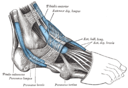
The mucous sheaths of the tendons around the ankle. Lateral aspect. (Extensor dig. longus labeled at upper right.) Latin musculus extensor digitorum longus Gray's subject #129 481 Origin Anterior lateral condyle of tibia, anterior shaft of fibula and superior ¾ of interosseous membrane Insertion Dorsal surface; middle and distal phalanges of lateral four digits Artery anterior tibial artery Nerve peroneal nerve Actions extension of toes and ankle Antagonist Flexor digitorum longus, Flexor digitorum brevis The Extensor digitorum longus is a pennate muscle, situated at the lateral part of the front of the leg.
Contents
Origin and insertion
It arises from the lateral condyle of the tibia; from the upper three-fourths of the anterior surface of the body of the fibula; from the upper part of the interosseous membrane; from the deep surface of the fascia; and from the intermuscular septa between it and the Tibialis anterior on the medial, and the Peronæi on the lateral side. Between it and the Tibialis anterior are the upper portions of the anterior tibial vessels and deep peroneal nerve.
The muscle passes under the transverse and cruciate crural ligaments in company with the Peronæus tertius, and divides into four slips, which run forward on the dorsum of the foot, and are inserted into the second and third phalanges of the four lesser toes.
The tendons to the second, third, and fourth toes are each joined, opposite the metatarsophalangeal articulation, on the lateral side by a tendon of the Extensor digitorum brevis. The tendons are inserted in the following manner: each receives a fibrous expansion from the Interossei and Lumbricalis, and then spreads out into a broad aponeurosis, which covers the dorsal surface of the first phalanx: this aponeurosis, at the articulation of the first with the second phalanx, divides into three slips—an intermediate, which is inserted into the base of the second phalanx; and two collateral slips, which, after uniting on the dorsal surface of the second phalanx, are continued onward, to be inserted into the base of the third phalanx.
Variations
This muscle varies considerably in the modes of origin and the arrangement of its various tendons.
The tendons to the second and fifth toes may be found doubled, or extra slips are given off from one or more tendons to their corresponding metatarsal bones, or to the short extensor, or to one of the interosseous muscles.
A slip to the great toe from the innermost tendon has been found.
Additional images
-
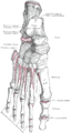
Bones of the right foot. Dorsal surface.
-
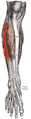
Muscles of the front of the leg.
-

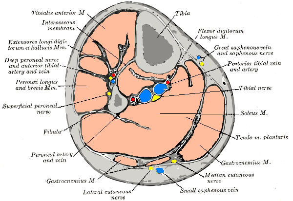
Cross-section through middle of leg.
-
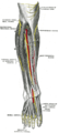
Deep nerves of the front of the leg.
See also
External links
- LUC exdl
- -590348211 at GPnotebook
- SUNY Labs 15:st-0401
- Extensor+digitorum+longus at eMedicine Dictionary
- PTCentral
This article was originally based on an entry from a public domain edition of Gray's Anatomy. As such, some of the information contained within it may be outdated.
List of muscles of lower limbs (TA A04.7, GA 4.465) ILIAC Region
/ ILIOPSOASBUTTOCKS THIGH /
compartmentsLEG/
Crus/
compartmentssuperficial · triceps surae (gastrocnemius, soleus, accessory soleus, Achilles tendon) · plantaris
deep · tarsal tunnel (flexor hallucis longus, flexor digitorum longus, tibialis posterior) · popliteusfibularis muscles (longus, brevis)FOOT DorsalPlantar1st layer (abductor hallucis, flexor digitorum brevis, abductor digiti minimi) · 2nd layer (quadratus plantae, lumbrical muscle) · 3rd layer (flexor hallucis brevis, adductor hallucis, flexor digiti minimi brevis) · 4th layer (dorsal interossei, plantar interossei)Categories:- Calf muscles
-


Wikimedia Foundation. 2010.