- DNA mismatch repair
-
DNA mismatch repair is a system for recognizing and repairing erroneous insertion, deletion and mis-incorporation of bases that can arise during DNA replication and recombination, as well as repairing some forms of DNA damage.[1][2]
Mismatch repair is strand-specific. During DNA synthesis the newly synthesised (daughter) strand will include errors commonly. In order to do this the mismatch repair machinery distinguishes the newly synthesised strand from the template (parental). In gram-negative bacteria transient hemimethylation distinguishes the strands (the parental is methylated and daughter is not). In other prokaryotes and eukaryotes the exact mechanism is not clear. It is suspected that in eukaryotes, newly synthesized lagging-strand DNA transiently contains nicks (before being sealed by DNA ligase) and provides a signal that directs mismatch proofreading systems to the appropriate strand. This implies that these nicks must be present in the leading strand, but it is unclear how.
Any mutational event that disrupts the superhelical structure of DNA carries with it the potential to compromise the genetic stability of a cell. The fact that the damage detection and repair systems are as complex as the replication machinery itself highlights the importance evolution has attached to DNA fidelity.
Examples of mismatched bases include a G/T or A/C pairing (see DNA repair). Mismatches are commonly due to tautomerization of bases during synthesis[citation needed]. The damage is repaired by recognition of the deformity caused by the mismatch, determining the template and non-template strand, and excising the wrongly incorporated base and replacing it with the correct nucleotide. The removal process involves more than just the mismatched nucleotide itself. A few or up to thousands of base pairs of the newly synthesized DNA strand can be removed.
Contents
Mismatch repair proteins
DNA mismatch repair protein, C-terminal domain 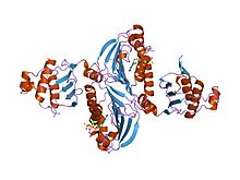
hpms2-atpgs Identifiers Symbol DNA_mis_repair Pfam PF01119 Pfam clan CL0329 InterPro IPR013507 PROSITE PDOC00057 SCOP 1bkn Available protein structures: Pfam structures PDB RCSB PDB; PDBe PDBsum structure summary Mismatch repair is a highly conserved process from prokaryotes to eukaryotes. The first evidence for mismatch repair was obtained from S. pneumoniae (the hexA and hexB genes). Subsequent work on E. coli has identified a number of genes that, when mutationally inactivated, cause hypermutable strains. The gene products are therefore called the "Mut" proteins, and are the major active components of the mismatch repair system. Three of these proteins are essential in detecting the mismatch and directing repair machinery to it; MutS, MutH and MutL (MutS is a homologue of HexA and MutL of HexB).
MutS forms a dimer (MutS2) that recognises the mismatched base on the daughter strand and binds the mutated DNA. MutH binds at hemimethylated sites along the daughter DNA, but its action is latent, being activated only upon contact by a MutL dimer (MutL2) which binds the MutS-DNA complex and acts as a mediator between MutS2 and MutH, activating the latter. The DNA is looped out to search for the nearest d(GATC) methylation site to the mismatch, which could be up to 1kb away. Upon activation by the MutS-DNA complex, MutH nicks the daughter strand near the hemimethylated site and recruits a UvrD helicase (DNA Helicase II) to separate the two strands with a specific 3' to 5' polarity. The entire MutSHL complex then slides along the DNA in the direction of the mismatch, liberating the strand to be excised as it goes. An exonuclease trails the complex and digests the ss-DNA tail. The exonuclease recruited is dependent on which side of the mismatch MutH incises the strand – 5’ or 3’. If the nick made by MutH is on the 5’ end of the mismatch, either RecJ or ExoVII (both 5’ to 3’ exonucleases) is used. If however the nick is on the 3’ end of the mismatch, ExoI (a 3' to 5' enzyme) is used.
The entire process ends past the mismatch site - i.e. both the site itself and its surrounding nucleotides are fully excised. The single-stranded gap created by the exonuclease can then be repaired by DNA Polymerase III (assisted by single-strand binding protein), which uses the other strand as a template, and finally sealed by DNA ligase. Dam methylase then rapidly methylates the daughter strand.
MutS homologs
When bound, the MutS2 dimer bends the DNA helix and shields approximately 20 base pairs. It has weak ATPase activity, and binding of ATP leads to the formation of tertiary structures on the surface of the molecule. The crystal structure of MutS reveals that it is exceptionally asymmetric, and while its active conformation is a dimer, only one of the two halves interact with the mismatch site.
In eukaryotes, MutS homologs form two major heterodimers: Msh2/Msh6 (MutSα) and Msh2/Msh3 (MutSβ). The MutSα pathway is involved primarily in base substitution and small loop mismatch repair. The MutSβ pathway is also involved in small loop repair, in addition to large loop (~10 nucleotide loops) repair. However, MutSβ does not repair base substitutions.
MutL homologs
MutL also has weak ATPase activity (it uses ATP for purposes of movement). It forms a complex with MutS and MutH, increasing the MutS footprint on the DNA.
However, the processivity (the distance the enzyme can move along the DNA before dissociating) of UvrD is only ~40–50bp. Because the distance between the nick created by MutH and the mismatch can average ~600 bp, if there isn't another UvrD loaded the unwound section is then free to re-anneal to its complementary strand, forcing the process to start over. However, when assisted by MutL, the rate of UvrD loading is greatly increased. While the processivity (and ATP utilisation) of the individual UvrD molecules remains the same, the total effect on the DNA is boosted considerably; the DNA has no chance to re-anneal, as each UvrD unwinds 40-50 bp of DNA, dissociates, and then is immediately replaced by another UvrD, repeating the process. This exposes large sections of DNA to exonuclease digestion, allowing for quick excision(and later replacement) of the incorrect DNA.
Eukaryotes have MutL homologs designated Mlh1 and Pms1. They form a heterodimer which mimics MutL in E. coli. The human homologue of prokaryotic MutL has three forms designated as MutLα, MutLβ and MutLγ. The MutLα complex is made of two subunits MLH1 and PMS2, the MutLβ heterodimer is made of MLH1 and PMS1, while MutLγ is made of MLH1 and MLH3. MutLα acts as the matchmaker or facilitator, coordinating events in mismatch repair. It has recently been shown to be a DNA endonuclease that introduces strand breaks in DNA upon activation by mismatch and other required proteins, MutSa and PCNA. These strand interruptions serve as entry points for an exonuclease activity that removes mismatched DNA. Roles played by MutLβ and MutLγ in mismatch repair are less well understood.
MutH: an endonuclease present in E. coli and Salmonella
MutH is a very weak endonuclease that is activated once bound to MutL (which itself is bound to MutS). It nicks unmethylated DNA and the unmethylated strand of hemimethylated DNA but does not nick fully methylated DNA. It has been experimentally shown that mismatch repair is random if neither strand is methylated. These behaviours led to the proposal that MutH determines which strand contains the mismatch. MutH has no eukaryotic homolog. Its endonuclease function is taken up by MutL homologs, which have some specialized 5'-3' exonuclease activity. The strand bias for removing mismatches from the newly synthesized daughter strand in eukaryotes may be provided by the free 3’ ends of Okazaki fragments in the new strand created during replication.
β-sliding clamp/PCNA
PCNA and the β-sliding clamp associate with MutSα/β and MutS, respectively. Although initial reports suggested that the PCNA-MutSα complex may enhance mismatch recognition,[3] it has been recently demonstrated[4] that there is no apparent change in affinity of MutSα for a mismatch in the presence or absence of PCNA. Furthermore, mutants of MutSα that are unable to interact with PCNA in vitro exhibit the capacity to carry out mismatch recognition and mismatch excision to near wild type levels. Curiously, such mutants are defective in the repair reaction directed by a 5' strand break, suggesting for the first time MutSα function in a post-excision step of the reaction.
Defects in mismatch repair
Mutations in the human homologues of the Mut proteins affect genomic stability, which can result in microsatellite instability (MI). MI is implicated in most human cancers. Specifically the overwhelming majority of hereditary nonpolyposis colorectal cancers (HNPCC) are attributed to mutations in the genes encoding the MutS and MutL homologues MSH2 and MLH1 respectively, which allows them to be classified as tumour suppressor genes. A subtype of HNPCC is known as Muir-Torre Syndrome (MTS) which is associated with skin tumors.
See also
References
- ^ Iyer R, Pluciennik A, Burdett V, Modrich P (2006). "DNA mismatch repair: functions and mechanisms". Chem Rev 106 (2): 302–23. doi:10.1021/cr0404794. PMID 16464007.
- ^ Larrea AA, Lujan SA, Kunkel TA (2010). "DNA mismatch repair". Cell 141 (4): 730. doi:10.1016/j.cell.2010.05.002. PMID 20478261.
- ^ Flores-Rozas H, Clark D, Kolodner RD (2000). "Proliferating cell nuclear antigen and Msh2p-Msh6p interact to form an active mispair recognition complex". Nature Genetics 26 (3): 375–8. doi:10.1038/81708. PMID 11062484.
- ^ Iyer RR, Pohlhaus TJ, Chen S, Hura GL, Dzantiev L, Beese LS, Modrich P (2008). "The MutSalpha-proliferating cell nuclear antigen interaction in human DNA mismatch repair". Journal of Biological Chemistry 283 (19): 13310–9. doi:10.1074/jbc.M800606200. PMC 2423938. PMID 18326858. http://www.pubmedcentral.nih.gov/articlerender.fcgi?tool=pmcentrez&artid=2423938.
Further reading
- Hsieh P, Yamane K (2008). "DNA mismatch repair: Molecular mechanism, cancer, and ageing". Mech Ageing Dev 129 (7-8): 391–407. doi:10.1016/j.mad.2008.02.012. PMC 2574955. PMID 18406444. http://www.pubmedcentral.nih.gov/articlerender.fcgi?tool=pmcentrez&artid=2574955.
- Iyer R, Pluciennik A, Burdett V, Modrich P (2006). "DNA mismatch repair: functions and mechanisms". Chem Rev 106 (2): 302–23. doi:10.1021/cr0404794. PMID 16464007.
- Joseph N, Duppatla V, Rao DN (2006). "Prokaryotic DNA mismatch repair". Prog. Nucleic Acid Res. Mol. Biol. 81: 1–49. doi:10.1016/S0079-6603(06)81001-9. PMID 16891168.
- Yang W (2000). "Structure and function of mismatch repair proteins". Mutat Res 460 (3-4): 245–56. PMID 10946232.
- Griffith, Wessler, Lewontin, Gelbart, Suzuki, Miller, Introduction to Genetic Analysis, 8th Edition, W.H. Freeman and Company, ISBN 0-7167-4939-4
- Thomas A.Kunkel and Dorothy A. Erie,(2005), DNA Mismatch Repair, Annu Rev.Biochem,74:681-710
- Errol C.Friedberg, Graham C. Walker, Wolfram Siede, Richard D. Wood, Roger A. Schultz, Tom Ellenberger, DNA repair and Mutagenesis, 2nd edition, ASM press, ISBN 1-55581-319-4
External links
Excision repair Other forms of repair Transcription-coupled repair (ERCC6, ERCC8) • DNA mismatch repair (MLH1, MSH2) • Homology directed repair • Non-homologous end joining (Ku) • Microhomology-mediated end joining • Postreplication repair • Photolyase (CRY1, CRY2)Other/ungrouped proteins Regulation Other/ungrouped 8-Oxoguanine • Adaptive response • Meiotic recombination checkpoint • RecF pathway
DNA helicase: BLM · WRN
FANC proteins: core protein complex (FANCA, FANCB, FANCC, FANCE, FANCF, FANCG, FANCL, FANCM), FANCD1, FANCD2, FANCI, FANCJ, FANCNCategories:

Wikimedia Foundation. 2010.