- Cardiac dysrhythmia
-
"Arrhythmia" redirects here. It is not to be confused with Erythema.
Cardiac dysrhythmia Classification and external resources 
Ventricular fibrillation (V-Fib or VF) an example of cardiac arrhythmia.ICD-10 I47 - I49 ICD-9 427 DiseasesDB 15206 MedlinePlus 001101 MeSH D001145 Cardiac dysrhythmia (also known as arrhythmia and irregular heartbeat) is a term for any of a large and heterogeneous group of conditions in which there is abnormal electrical activity in the heart. The heart beat may be too fast or too slow, and may be regular or irregular.
Some arrhythmias are life-threatening medical emergencies that can result in cardiac arrest and sudden death. Others cause symptoms such as an abnormal awareness of heart beat (palpitations), and may be merely annoying. These palpitations have also been known to be caused by atrial/ventricular fibrillation, wire faults, and other technical or mechanical issues in cardiac pacemakers/defibrillators. Still others may not be associated with any symptoms at all, but may predispose the patient to potentially life threatening stroke or embolism.
Some arrhythmias are very minor and can be regarded as normal variants. In fact, most people will on occasion feel their heart skip a beat, or give an occasional extra strong beat; neither of these is usually a cause for alarm.[1]
Proarrhythmia is a new or more frequent occurrence of pre-existing arrhythmias, paradoxically precipitated by antiarrhythmic therapy, which means it is a side effect associated with the administration of some existing antiarrhythmic drugs, as well as drugs for other indications. In other words, it is a tendency of antiarrhythmic drugs to facilitate emergence of new arrhythmias.
The term sinus arrhythmia refers to a normal phenomenon of mild acceleration and slowing of the heart rate that occurs with breathing in and out. It is usually quite pronounced in children, and steadily decreases with age. This can also be present during meditation breathing exercises that involve deep inhaling and breath holding patterns.
Contents
Classification
Arrhythmia may be classified by rate (normal, tachycardia, bradycardia), or mechanism (automaticity, reentry, fibrillation).
It is also appropriate to classify by site of origin:
Atrial
- Premature Atrial Contractions (PACs)
- Wandering Atrial Pacemaker
- Multifocal atrial tachycardia
- Atrial flutter
- Atrial fibrillation (Afib)
Junctional arrhythmias
- Supraventricular tachycardia (SVT)
- AV nodal reentrant tachycardia is the most common cause of Paroxysmal Supra-ventricular Tachycardia (PSVT)
- Junctional rhythm
- Junctional tachycardia
- Premature junctional complex
Ventricular
- Premature Ventricular Contractions (PVC) sometimes called Ventricular Extra Beats (VEBs)
- Premature Ventricular beats occurring after every normal beat are termed "ventricular bigeminy"
- PVCs that occur at intervals of 2 normal beats to 1 PVC are termed "PVCs in trigeminy"
- Three premature ventricular grouped together is termed a "run of PVCs"; runs lasting longer than three beats are generally referred to as ventricular tachycardia
- Accelerated idioventricular rhythm
- Monomorphic Ventricular tachycardia
- Polymorphic ventricular tachycardia
- Ventricular fibrillation
Heart blocks
These are also known as AV blocks, because the vast majority of them arise from pathology at the atrioventricular node. They are the most common causes of bradycardia:
- First degree heart block, which manifests as PR prolongation
- Second degree heart block
- Type 1 Second degree heart block, also known as Mobitz I or Wenckebach
- Type 2 Second degree heart block, also known as Mobitz II
- Third degree heart block, also known as complete heart block.
SADS
SADS, or sudden arrhythmic death syndrome, is a term used to describe sudden death due to cardiac arrest brought on by an arrhythmia in the absence of any structural heart disease on autopsy. The most common cause of sudden death in the US is coronary artery disease.[citation needed] Approximately 300,000 people die suddenly of this cause every year in the US.[citation needed] SADS occurs from other causes. There are many inherited conditions and heart diseases that can affect young people and subsequently cause sudden death. Many of these victims have no symptoms before dying suddenly.[citation needed]
Causes of SADS in young people include viral myocarditis, long QT syndrome, Brugada syndrome, Catecholaminergic polymorphic ventricular tachycardia, hypertrophic cardiomyopathy and arrhythmogenic right ventricular dysplasia.[citation needed]
Signs and symptoms
The term cardiac arrhythmia covers a very large number of very different conditions.
The most common symptom of arrhythmia is an abnormal awareness of heartbeat, called palpitations. These may be infrequent, frequent, or continuous. Some of these arrhythmias are harmless (though distracting for patients) but many of them predispose to adverse outcomes.
Some arrhythmias do not cause symptoms, and are not associated with increased mortality. However, some asymptomatic arrhythmias are associated with adverse events. Examples include a higher risk of blood clotting within the heart and a higher risk of insufficient blood being transported to the heart because of weak heartbeat. Other increased risks are of embolisation and stroke, heart failure and sudden cardiac death.
If an arrhythmia results in a heartbeat that is too fast, too slow or too weak to supply the body's needs, this manifests as a lower blood pressure and may cause lightheadedness or dizziness, or fainting.
Some types of arrhythmia result in cardiac arrest, or sudden death.
Medical assessment of the abnormality using an electrocardiogram is one way to diagnose and assess the risk of any given arrhythmia.
Differential diagnosis
Normal electrical activity
Main article: Electrical conduction system of the heartEach heart beat originates as an electrical impulse from a small area of tissue in the right atrium of the heart called the sinus node or Sino-atrial node or SA node. The impulse initially causes both atria to contract, then activates the atrioventricular (or AV) node which is normally the only electrical connection between the atria and the ventricles (main pumping chambers). The impulse then spreads through both ventricles via the Bundle of His and the Purkinje fibres causing a synchronised contraction of the heart muscle and, thus, the pulse.
In adults the normal resting heart rate ranges from 60 to 80 beats per minute. The resting heart rate in children is much faster.
Bradycardias
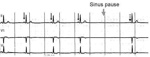
 Normal sinus rhythm, with solid black arrows pointing to normal P waves representative of normal sinus node function, followed by a pause in sinus node activity (resulting in a transient loss of heart beats). Note that the P wave that disrupts the pause (indicated by the dashed arrow) does not look like the previous (normal) P waves — this last P wave is arising from a different part of the atrium, representing an escape rhythm.
A slow rhythm (less than 60 beats/min), is labelled bradycardia. This may be caused by a slowed signal from the sinus node (sinus bradycardia), a pause in the normal activity of the sinus node (sinus arrest), or by blocking of the electrical impulse on its way from the atria to the ventricles (AV block or heart block). Heart block comes in varying degrees and severity. It may be caused by reversible poisoning of the AV node (with drugs that impair conduction) or by irreversible damage to the node. Bradycardias may also be present in the normally functioning heart of endurance athletes or other well-conditioned persons.
Tachycardias
In adults and children over 15, resting heart rate faster than 100 beats/minute is labelled tachycardia. Tachycardia may result in palpitation; however, tachycardia is not necessarily an arrhythmia. Increased heart rate is a normal response to physical exercise or emotional stress. This is mediated by the sympathetic nervous system on the sinus node and called sinus tachycardia. Other things that increase sympathetic nervous system activity in the heart include ingested or injected substances, such as caffeine or amphetamines, and an overactive thyroid gland (hyperthyroidism).
Tachycardia that is not sinus tachycardia usually results from the addition of abnormal impulses to the normal cardiac cycle. Abnormal impulses can begin by one of three mechanisms: automaticity, reentry or triggered activity. A specialised form of re-entry problem is termed fibrillation.
Automaticity
Automaticity refers to a cardiac muscle cell firing off an impulse on its own. All of the cells in the heart have the ability to initiate an action potential; however, only some of these cells are designed to routinely trigger heart beats. These cells are found in the conduction system of the heart and include the SA node, AV node, Bundle of His and Purkinje fibers. The sinoatrial node is a single specialized location in the atrium which has a higher automaticity (a faster pacemaker) than the rest of the heart and, therefore, is usually responsible for setting the heart rate and initiating each heart beat.
Any part of the heart that initiates an impulse without waiting for the sinoatrial node is called an ectopic focus and is, by definition, a pathological phenomenon. This may cause a single premature beat now and then, or, if the ectopic focus fires more often than the sinoatrial node, it can produce a sustained abnormal rhythm. Rhythms produced by an ectopic focus in the atria, or by the atrioventricular node, are the least dangerous dysrhythmias; but they can still produce a decrease in the heart's pumping efficiency, because the signal reaches the various parts of the heart muscle with different timing to usual and can be responsible for poorly coordinated contraction.
Conditions that increase automaticity include sympathetic nervous system stimulation and hypoxia. The resulting heart rhythm depends on where the first signal begins: If it is the sinoatrial node, the rhythm remains normal but rapid; if it is an ectopic focus, many types of dysrhythmia may ensue.
Re-entry
Re-entry arrhythmias occur when an electrical impulse recurrently travels in a tight circle within the heart, rather than moving from one end of the heart to the other and then stopping.[2] Every cardiac cell is able to transmit impulses in every direction but will only do so once within a short time. Normally, the action potential impulse will spread through the heart quickly enough that each cell will only respond once. However, if conduction is abnormally slow in some areas (for example in heart damage) so the myocardial cells are unable to activate the fast sodium channel, part of the impulse will arrive late and potentially be treated as a new impulse. Depending on the timing, this can produce a sustained abnormal circuit rhythm. Re-entry circuits are responsible for atrial flutter, most paroxysmal supraventricular tachycardia, and dangerous ventricular tachycardia. These types of re-entry circuits are different from WPW syndromes in which the real pathways existed.
Fibrillation
When an entire chamber of the heart is involved in a multiple micro-reentry circuits and, therefore, quivering with chaotic electrical impulses, it is said to be in fibrillation.
Fibrillation can affect the atrium (atrial fibrillation) or the ventricle (ventricular fibrillation); ventricular fibrillation is imminently life-threatening.
- Atrial fibrillation affects the upper chambers of the heart, known as the atria. Atrial fibrillation may be due to serious underlying medical conditions and should be evaluated by a physician. It is not typically a medical emergency.
- Ventricular fibrillation occurs in the ventricles (lower chambers) of the heart; it is always a medical emergency. If left untreated, ventricular fibrillation (VF, or V-fib) can lead to death within minutes. When a heart goes into V-fib, effective pumping of the blood stops. V-fib is considered a form of cardiac arrest. An individual suffering from it will not survive unless cardiopulmonary resuscitation (CPR) and defibrillation are provided immediately.
CPR can prolong the survival of the brain in the lack of a normal pulse, but defibrillation is the only intervention that can restore a healthy heart rhythm. Defibrillation is performed by applying an electric shock to the heart, which resets the cells, permitting a normal beat to re-establish itself.
Triggered beats
Triggered beats occur when problems at the level of the ion channels in individual heart cells result in abnormal propagation of electrical activity and can lead to sustained abnormal rhythm. They are relatively rare and can result from the action of anti-arrhythmic drugs.
Diagnostic approach
Cardiac dysrhythmias are often first detected by simple but nonspecific means: auscultation of the heartbeat with a stethoscope, or feeling for peripheral pulses. These cannot usually diagnose specific dysrhythmias, but can give a general indication of the heart rate and whether it is regular or irregular. Not all the electrical impulses of the heart produce audible or palpable beats; in many cardiac arrhythmias, the premature or abnormal beats do not produce an effective pumping action and are experienced as "skipped" beats.
The simplest specific diagnostic test for assessment of heart rhythm is the electrocardiogram (abbreviated ECG or EKG). A Holter monitor is an EKG recorded over a 24-hour period, to detect dysrhythmias that may happen briefly and unpredictably throughout the day.
A more advanced study of the heart's electrical activity can be performed to assess the source of the aberrant heart beats. This can be accomplished in an Electrophysiology study. A minimally invasive procedure that uses a catheter to "listen" to the electrical activity from within the heart, additionally if the source of the arrhythmias is found, often the abnormal cells can be ablated and the arrhythmia can be permanently corrected.
Management
The method of cardiac rhythm management depends firstly on whether or not the affected person is stable or unstable. Treatments may include physical maneuvers, medications, electricity conversion, or electro or cryo cautery.
Physical maneuvers
A number of physical acts can increase parasympathetic nervous supply to the heart, resulting in blocking of electrical conduction through the AV node. This can slow down or stop a number of arrhythmias that originate above or at the AV node (see main article: supraventricular tachycardias). Parasympathetic nervous supply to the heart is via the vagus nerve, and these maneuvers are collectively known as vagal maneuvers.
Antiarrhythmic drugs
Main article: Antiarrhythmic agentsThere are many classes of antiarrhythmic medications, with different mechanisms of action and many different individual drugs within these classes. Although the goal of drug therapy is to prevent arrhythmia, nearly every antiarrhythmic drug has the potential to act as a pro-arrhythmic, and so must be carefully selected and used under medical supervision.
Other drugs
A number of other drugs can be useful in cardiac arrhythmias.
Several groups of drugs slow conduction through the heart, without actually preventing an arrhythmia. These drugs can be used to "rate control" a fast rhythm and make it physically tolerable for the patient.
Some arrhythmias promote blood clotting within the heart, and increase risk of embolus and stroke. Anticoagulant medications such as warfarin and heparins, and anti-platelet drugs such as aspirin can reduce the risk of clotting.
Electricity
Dysrhythmias may also be treated electrically, by applying a shock across the heart — either externally to the chest wall, or internally to the heart via implanted electrodes.
Cardioversion is either achieved pharmacologically or via the application of a shock synchronised to the underlying heartbeat. It is used for treatment of supraventricular tachycardias. In elective cardioversion, the recipient is usually sedated or lightly anesthetized for the procedure.
Defibrillation differs in that the shock is not synchronised. It is needed for the chaotic rhythm of ventricular fibrillation and is also used for pulseless ventricular tachycardia. Often, more electricity is required for defibrillation than for cardioversion. In most defibrillation, the recipient has lost consciousness so there is no need for sedation.
Defibrillation or cardioversion may be accomplished by an implantable cardioverter-defibrillator (ICD).
Electrical treatment of dysrhythmia also includes cardiac pacing. Temporary pacing may be necessary for reversible causes of very slow heartbeats, or bradycardia, (for example, from drug overdose or myocardial infarction). A permanent pacemaker may be placed in situations where the bradycardia is not expected to recover.
Electrical cautery
Some cardiologists further sub-specialise into electrophysiology. In specialised catheter laboratories, they use fine probes inserted through the blood vessels to map electrical activity from within the heart. This allows abnormal areas of conduction to be located very accurately, and subsequently destroyed with heat, cold, electrical or laser probes.
This may be completely curative for some forms of arrhythmia, but for others, the success rate remains disappointing. AV nodal reentrant tachycardia is often curable. Atrial fibrillation can also be treated with this technique (e.g. pulmonary vein isolation), but the results are less reliable.
See also
- Palpitation
- Clinical cardiac electrophysiology
- Antiarrhythmic agents
- Artificial pacemaker
- Electrical conduction system of the heart
- Implantable cardioverter-defibrillator
- Electrophysiology Study
- Radiofrequency ablation
References
- ^ familydoctor.org
- ^ Allessie MA, Bonke FI, Schopman FJ (August 1976). "Circus movement in rabbit atrial muscle as a mechanism of tachycardia. II. The role of nonuniform recovery of excitability in the occurrence of unidirectional block, as studied with multiple microelectrodes". Circ. Res. 39 (2): 168–77. PMID 939001.
External links
Categories:- Cardiac dysrhythmia
- Medical emergencies

Wikimedia Foundation. 2010.