- Commissural fiber
-
Brain: Commissural fiber 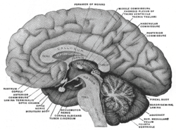
Mesal aspect of a brain sectioned in the median sagittal plane. Latin fibra commissuralis, fibrae commissurales telencephali Gray's subject #189 843 NeuroNames ancil-221 Code TA A14.1.00.017 The commissural fibers or transverse fibers connect the two hemispheres of the brain. They include:
- the transverse fibers of the corpus callosum
- the anterior commissure
- the posterior commissure
- the lyra or hippocampal commissure.
In contrast to commissural fibers, association fibers connect regions within the same hemisphere of the brain. Projection fibers connect the lobe to other parts of the brain and the spinal cord.
External links
This article was originally based on an entry from a public domain edition of Gray's Anatomy. As such, some of the information contained within it may be outdated.
Histology: nervous tissue (TA A14, GA 9.849, TH H2.00.06, H3.11) CNS GeneralGrey matter · White matter (Projection fibers · Association fiber · Commissural fiber · Lemniscus · Funiculus · Fasciculus · Decussation · Commissure) · meningesOtherPNS GeneralPosterior (Root, Ganglion, Ramus) · Anterior (Root, Ramus) · rami communicantes (Gray, White) · Autonomic ganglion (Preganglionic nerve fibers · Postganglionic nerve fibers)Myelination: Schwann cell (Neurolemma, Myelin incisure, Myelin sheath gap, Internodal segment)
Satellite glial cellNeurons/
nerve fibersPartsPerikaryon (Axon hillock)
Axon (Axon terminals, Axoplasm, Axolemma, Neurofibril/neurofilament)
Dendrite (Nissl body, Dendritic spine, Apical dendrite/Basal dendrite)TypesGSA · GVA · SSA · SVA
fibers (Ia, Ib or Golgi, II or Aβ, III or Aδ or fast pain, IV or C or slow pain)GSE · GVE · SVE
Upper motor neuron · Lower motor neuron (α motorneuron, γ motorneuron, β motorneuron)Termination SynapseHuman brain, cerebrum, Interior of the cerebral hemispheres, white matter: commissural fibers and septum (TA A14.1.09.241–271, 569–571, GA 9.828, 838–840) Corpus callosum Genu · Splenium · Tapetum · Rostrum
Archicortex: Indusium griseumLamina terminalis Fornix Septum pellucidum Categories:- Neuroscience stubs
- Nervous system

Wikimedia Foundation. 2010.