- Muscular system
-
Muscular system Latin = systema musculare
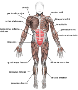
Muscles anterior labeled 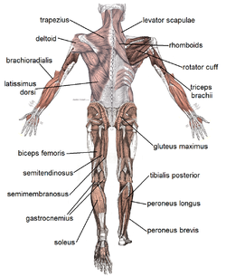
Muscle posterior labeled The muscular system is the anatomical system of a species that allows it to move. The muscular system in vertebrates is controlled through the nervous system, although some muscles (such as the cardiac muscle) can be completely autonomous.
Contents
Muscles
Main article: MuscleThere are three distinct types of muscles: skeletal muscles, cardiac or heart muscles, and smooth (non-striated) muscles. Muscles provide strength, balance, posture, movement and heat for the body to keep warm.
Upon stimulation by an action potential, skeletal muscles perform a coordinated contraction by shortening each sarcomere. The best proposed model for understanding contraction is the sliding filament model of muscle contraction. Actin and myosin fibers overlap in a contractile motion towards each other. Myosin filaments have club-shaped heads that project toward the actin filaments.
Larger structures along the myosin filament called myosin heads are used to provide attachment points on binding sites for the actin filaments. The myosin heads move in a coordinated style, they swivel toward the center of the sarcomere, detach and then reattach to the nearest active site of the actin filament. This is called a rachet type drive system. This process consumes large amounts of adenosine triphosphate (ATP).
Energy for this comes from ATP, the energy source of the cell. ATP binds to the cross bridges between myosin heads and actin filaments. The release of energy powers the swiveling of the myosin head. Muscles store little ATP and so must continuously recycle the discharged adenosine diphosphate molecule (ADP) into ATP rapidly. Muscle tissue also contains a stored supply of a fast acting recharge chemical, creatine phosphate which can assist initially producing the rapid regeneration of ADP into ATP.
Calcium ions are required for each cycle of the sarcomere. Calcium is released from the sarcoplasmic reticulum into the sarcomere when a muscle is stimulated to contract. This calcium uncovers the actin binding sites. When the muscle no longer needs to contract, the calcium ions are pumped from the sarcomere and back into storage in the sarcoplasmic reticulum.
Anatomy
Main article: Table of muscles of the human bodyThere are approximately 639 skeletal muscles in the human body.
The following are some major muscles[1] and their basic features:
Aerobic and anaerobic muscle activity
At rest, the body produces the majority of its ATP aerobically in the mitochondria[2] without producing lactic acid or other fatiguing byproducts.[3] During exercise, the method of ATP production varies depending on the fitness of the individual as well as the duration, and intensity of exercise. At lower activity levels, when exercise continues for a long duration (several minutes or longer), energy is produced aerobically by combining oxygen with carbohydrates and fats stored in the body. Activity that is higher in intensity, with possible duration decreasing as intensity increases, ATP production can switch to anaerobic pathways, such as the use of the creatine phosphate and the phosphagen system or anaerobic glycolysis. Aerobic ATP production is biochemically much slower and can only be used for long-duration, low intensity exercise, but produces no fatiguing waste products that can not be removed immediately from sarcomere and body and results in a much greater number of ATP molecules per fat or carbohydrate molecule. Aerobic training allows the oxygen delivery system to be more efficient, allowing aerobic metabolism to begin quicker.[3] Anaerobic ATP production produces ATP much faster and allows near-maximal intensity exercise, but also produces significant amounts of lactic acid which render high intensity exercise unsustainable for greater than several minutes.[3] The phosphagen system is also anaerobic, allows for the highest levels of exercise intensity, but intramuscular stores of phosphocreatine are very limited and can only provide energy for exercises lasting up to ten seconds. Recovery is very quick, with full creatine stores regenerated within five minutes.[3]
Cardiac muscle
Main article: Heart muscleHeart muscles are distinct from skeletal muscles because the muscle fibers are laterally connected to each other. Furthermore, just as with smooth muscles, they are not controlling themselves. Heart muscles are controlled by the sinus node influenced by the autonomic nervous system.
Smooth muscle
Main article: Smooth muscleSmooth muscles are controlled directly by the autonomic nervous system and are involuntary, meaning that they are incapable of being moved by conscious thought. Functions such as heart beat and lungs (which are capable of being willingly controlled, be it to a limited extent) are involuntary muscles but are not smooth muscles.
Control of muscle contraction
Neuromuscular junctions are the focal point where a motor neuron attaches to a muscle. Acetylcholine, (a neurotransmitter used in skeletal muscle contraction) is released from the axon terminal of the nerve cell when an action potential reaches the microscopic junction, called a synapse. A group of chemical messengers cross the synapse and stimulate the formation of electrical changes, which are produced in the muscle cell when the acetylcholine binds to receptors on its surface. Calcium is released from its storage area in the cell's sarcoplasmic reticulum. An impulse from a nerve cell causes calcium release and brings about a single, short muscle contraction called a muscle twitch. If there is a problem at the neuromuscular junction, a very prolonged contraction may occur, tetanus. Also, a loss of function at the junction can produce paralysis.
Skeletal muscles are organized into hundreds of motor units, each of which involves a motor neuron, attached by a series of thin finger-like structures called axon terminals. These attach to and control discrete bundles of muscle fibers. A coordinated and fine tuned response to a specific circumstance will involve controlling the precise number of motor units used. While individual muscle units contract as a unit, the entire muscle can contract on a predetermined basis due to the structure of the motor unit. Motor unit coordination, balance, and control frequently come under the direction of the cerebellum of the brain. This allows for complex muscular coordination with little conscious effort, such as when one drives a car without thinking about the process.
See also
- Major systems of the human body
Notes
- ^ List of major muscles of the human body
- ^ ABERCROMBIE M; HICKMAN C.J; JOHNSON M.L, 1973, A Dictionary of Biology, Page 179, Middlesex (England), Baltimore (U.S.A), Ringwood (Australia): Penguin Books
- ^ a b c d "St Paul’s College Stage 2 EXERCISE PHYSIOLOGY Energy Systems Part 5" (ppt). http://intranet.spc.sa.edu.au/usermedia/curr/pe/ep5.ppt. Retrieved 2007-10-16.
References
Medical and Health Encyclopedia, chapter 1
External links
- GetBody Smart Muscle system tutorials and quizzes
- MedBio.info Use and formation of ATP in muscle
Human systems and organs TA 2–4:
MSBone (Carpus · Collar bone (clavicle) · Thigh bone (femur) · Fibula · Humerus · Mandible · Metacarpus · Metatarsus · Ossicles · Patella · Phalanges · Radius · Skull (cranium) · Tarsus · Tibia · Ulna · Rib · Vertebra · Pelvis · Sternum) · CartilageMuscular systemTA 5–11:
splanchnic/
viscusmostly
Thoracicmostly
AbdominopelvicDigestive system+
adnexaMouth (Salivary gland, Tongue) · upper GI (Oropharynx, Laryngopharynx, Esophagus, Stomach) · lower GI (Small intestine, Appendix, Colon, Rectum, Anus) · accessory (Liver, Biliary tract, Pancreas)TA 12–16 Blood
(Non-TA)General anatomy: systems and organs, regional anatomy, planes and lines, superficial axial anatomy, superficial anatomy of limbs Muscular system (TA A04.0, GA 4.361) Topics Types of muscles Other Unipennate muscle · Bipennate muscle · Origin · Insertion · Fascia (Superficial fascia, Deep fascia, Visceral fascia) · Tendon/Aponeurosis · Fascial compartmentCategories:


Wikimedia Foundation. 2010.