- Medial pterygoid muscle
-
Medial pterygoid 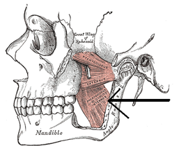
The Pterygoidei; the zygomatic arch and a portion of the ramus of the mandible have been removed. (Internus is visible at center bottom.) 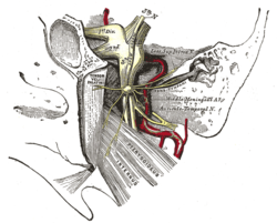
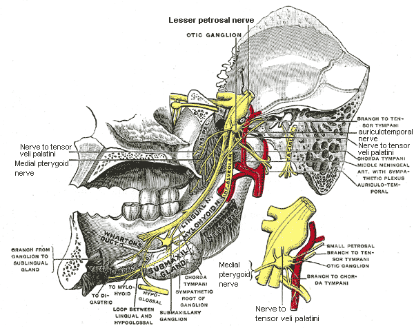
The otic ganglion and its branches. (Pterygoideus internus labeled at bottom right.) Latin musculus pterygoideus medialis, musculus pterygoideus internus Gray's subject #109 387 Origin deep head: medial side of lateral pterygoid plate behind the upper teeth
superficial head: pyramidal process of palatine bone and maxillary tuberosityInsertion medial angle of the mandible Artery pterygoid branches of maxillary artery Nerve mandibular nerve via nerve to medial pterygoid Actions elevates mandible, closes jaw, helps lateral pterygoids in moving the jaw from side to side MeSH Pterygoid+Muscles The medial pterygoid (or internal pterygoid muscle), is a thick, quadrilateral muscle of mastication.
The mandibular branch of the fifth cranial nerve, the trigeminal nerve, innervates the medial pterygoid muscle.
Contents
Origin and insertion
It consists of two heads.
- The bulk of the muscle arises as a deep head from just above the medial surface of the lateral pterygoid plate.
- The smaller, superficial head originates from the maxillary tuberosity and the pyramidal process of the palatine bone.
Its fibers pass downward, lateral, and posterior, and are inserted, by a strong tendinous lamina, into the lower and back part of the medial surface of the ramus and angle of the mandible, as high as the mandibular foramen. The insertion joins the masseter muscle to form a common tendinous sling which allows the medial pterygoid and masseter to be powerful elevators of the jaw.
Innervation
Like the lateral pterygoid, and all other muscles of mastication (apart from buccinator which is innervated by the facial nerve (VII)) the medial pterygoid is innervated by the anterior root (motor root) of the mandibular branch of the trigeminal nerve (V). Note: the buccinator muscle is not a muscle of mastication; instead it is classified as a facial muscle.
Actions
Given that the origin is on the medial side of the lateral pterygoid plate and the insertion is from the internal surface of the ramus of the mandible down to the angle of the mandible, its functions include:
- Elevation of the mandible (closes the jaw)
- Minor contribution to protrusion of the mandible
- Assistance in mastication
- Excursion of the mandible; contralateral excursion occurs with unilateral contraction.
Additional images
-
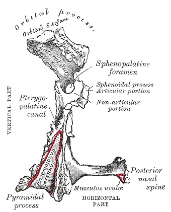
Left palatine bone. Posterior aspect. Enlarged.
-

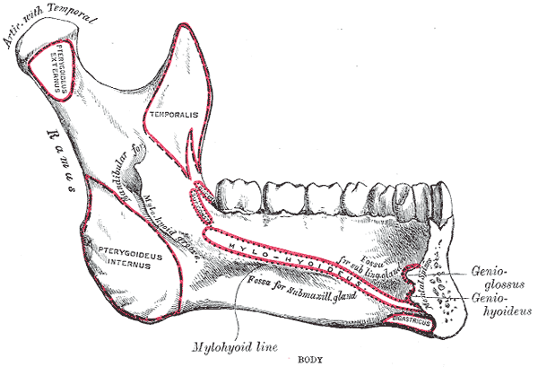
Mandible. Inner surface. Side view.
-
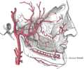
Plan of branches of internal maxillary artery.
-

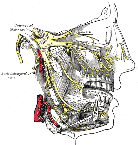
Distribution of the maxillary and mandibular nerves, and the submaxillary ganglion.
-

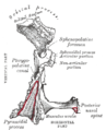
Mandibular division of trifacial nerve, seen from the middle line.
-
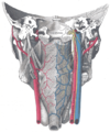
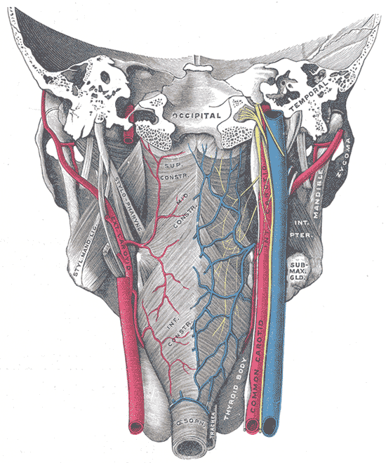
Muscles of the pharynx, viewed from behind, together with the associated vessels and nerves.
External links
- LUC mpte
- -1483079600 at GPnotebook
- Mnemonic at medicalmnemonics.com 70
- Roche Lexicon - illustrated navigator, at Elsevier 25420.000-1
This article was originally based on an entry from a public domain edition of Gray's Anatomy. As such, some of the information contained within it may be outdated.
List of muscles of head and neck: the head (TA A04.1, GA 4.378) Extraocular (CN III, IV, VI) oblique (inferior, superior) · rectus (superior, inferior, medial, lateral) · levator palpebrae superioris (superior tarsal)Mastication (CN V3) masseter · temporalis (sphenomandibularis) · pterygoid (lateral, medial)
fascia: Masseteric fascia · Temporal fascia · Deep portion: cementomaxillary tendon · Superficial portion: cementomandibular tendonFacial (CN VII) levator anguli oris · levator labii superioris · zygomaticus (major, minor)
orbicularis oris · risorius · buccinator
depressor anguli oris · depressor labii inferioris · mentalisPalate/fauces (CN IX, X, XI)
(except TVP=V3)veli palatini (tensor, levator) · musculus uvulae · palatopharyngeus (to pharynx) · palatoglossus (to tongue)Tongue (CN XII) extrinsic (genioglossus, hyoglossus/chondroglossus, styloglossus, and palatoglossus) · intrinsic (superior longitudinal, inferior longitudinal, transverse, vertical)Also it elevates mandible.
Categories:- Muscle stubs
- Muscles of the head and neck


Wikimedia Foundation. 2010.