- Golgi apparatus
-
This article is about the organelle. For the Phish song, see Junta (album).
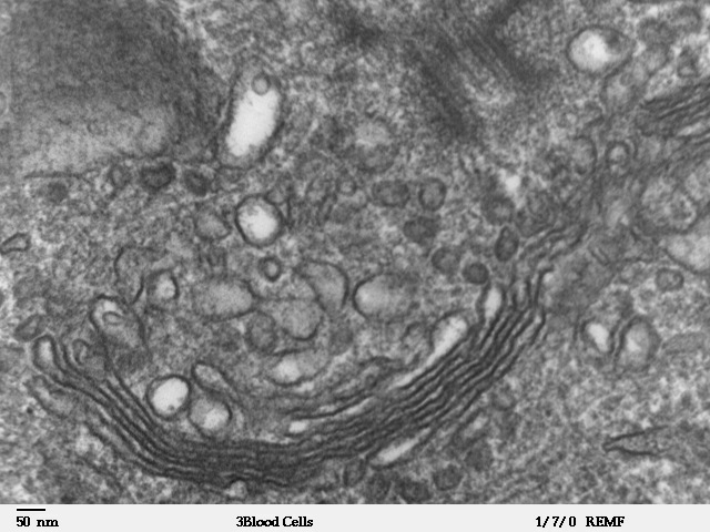
 Micrograph of Golgi apparatus, visible as a stack of semicircular black rings near the bottom. Numerous circular vesicles can be seen in proximity to the organelle
 Diagram of secretory process from endoplasmic reticulum (orange) to Golgi apparatus (pink). 1. Nuclear membrane; 2. Nuclear pore; 3. Rough endoplasmic reticulum (RER); 4. Smooth endoplasmic reticulum (SER); 5. Ribosome attached to RER; 6. Macromolecules; 7. Transport vesicles; 8. Golgi apparatus; 9. Cis face of Golgi apparatus; 10. Trans face of Golgi apparatus; 11. Cisternae of lipids
The Golgi apparatus (Golgi complex) is an organelle found in most eukaryotic cells.[1] It was identified in 1898 by the Italian physician Camillo Golgi, after whom the Golgi apparatus is named.[2]
It processes and packages proteins after their synthesis and before they make their way to their destination; it is particularly important in the processing of proteins for secretion. The Golgi apparatus forms a part of the cellular endomembrane system.
Contents
Discovery
Due to its fairly large size, the Golgi apparatus was one of the first organelles to be discovered and observed in detail. The apparatus was discovered in 1898 by Italian physician Camillo Golgi during an investigation of the nervous system.[2] After first observing it under his microscope, he termed the structure the internal reticular apparatus. The structure was then renamed after Golgi not long after the announcement of his discovery in 1898. However, some doubted the discovery at first, arguing that the appearance of the structure was merely an optical illusion created by the observation technique used by Golgi. With the development of modern microscopes in the 20th century, the discovery was confirmed.[3]
Structure
Found within the cytoplasm of both plant and animal cells, the Golgi is composed of stacks of membrane-bound structures known as cisternae (singular: cisterna). An individual stack is sometimes called a dictyosome (from Greek dictyon: net + soma: body),[4] especially in plant cells.[5] A mammalian cell typically contains 40 to 100 stacks.[6] Between four and eight cisternae are usually present in a stack; however, in some protists as many as sixty have been observed.[3] Each cisterna comprises a flat, membrane enclosed disc that includes special Golgi enzymes which modify or help to modify cargo proteins that travel through it.[7]
The cisternae stack has four functional regions: the cis-Golgi network, medial-Golgi, endo-Golgi, and trans-Golgi network. Vesicles from the endoplasmic reticulum (via the vesicular-tubular clusters) fuse with the network and subsequently progress through the stack to the trans Golgi network, where they are packaged and sent to the required destination. Each region contains different enzymes which selectively modify the contents depending on where they reside.[8] The cisternae also carry structural proteins important for their maintenance as flattened membranes which stack upon each other.[9]
Function
Cells synthesise a large number of different macromolecules. The Golgi apparatus is integral in modifying, sorting, and packaging these macromolecules for cell secretion[10] (exocytosis) or use within the cell.[11] It primarily modifies proteins delivered from the rough endoplasmic reticulum but is also involved in the transport of lipids around the cell, and the creation of lysosomes.[11] In this respect it can be thought of as similar to a post office; it packages and labels items which it then sends to different parts of the cell.
Enzymes within the cisternae are able to modify the proteins by addition of carbohydrates (glycosylation)[12] and phosphates (phosphorylation). In order to do so, the Golgi imports substances such as nucleotide sugars from the cytosol. These modifications may also form a signal sequence which determines the final destination of the protein. For example, the Golgi apparatus adds a mannose-6-phosphate label to proteins destined for lysosomes.
The Golgi plays an important role in the synthesis of proteoglycans, which are molecules present in the extracellular matrix of animals. It is also a major site of carbohydrate synthesis.[13] This includes the production of glycosaminoglycans (GAGs), long unbranched polysaccharides which the Golgi then attaches to a protein synthesised in the endoplasmic reticulum to form proteoglycans.[14] Enzymes in the Golgi polymerize several of these GAGs via a xylose link onto the core protein. Another task of the Golgi involves the sulfation of certain molecules passing through its lumen via sulfotranferases that gain their sulfur molecule from a donor called PAPs. This process occurs on the GAGs of proteoglycans as well as on the core protein. The level of sulfation is very important to the proteoglycans' signalling abilities as well as giving the proteoglycan its overall negative charge.[13]
The phosphorylation of molecules requires that ATP is imported into the lumen of the Golgi[15] and then utilised by resident kinases such as casein kinase 1 and casein kinase 2. One molecule that is phosphorylated in the Golgi is Apolipoprotein, which forms a molecule known as VLDL that is a constituent of blood serum. It is thought that the phosphorylation of these molecules is important to help aid in their sorting for secretion into the blood serum.[16]
The Golgi has a putative role in apoptosis, with several Bcl-2 family members localised there, as well as to the mitochondria. A newly characterized protein, GAAP (Golgi anti-apoptotic protein), almost exclusively resides in the Golgi and protects cells from apoptosis by an as-yet undefined mechanism.[17]
Vesicular transport
The vesicles that leave the rough endoplasmic reticulum are transported to the cis face of the Golgi apparatus, where they fuse with the Golgi membrane and empty their contents into the lumen. Once inside the lumen, the molecules are modified, then sorted for transport to their next destinations. The Golgi apparatus tends to be larger and more numerous in cells that synthesise and secrete large amounts of substances, for example, the plasma B cells and the antibody-secreting cells of the immune system have prominent Golgi complexes.
Those proteins destined for areas of the cell other than either the endoplasmic reticulum or Golgi apparatus are moved towards the trans face, to a complex network of membranes and associated vesicles known as the trans-Golgi network (TGN).[8] This area of the Golgi is the point at which proteins are sorted and shipped to their intended destinations by their placement into one of at least three different types of vesicles, depending upon the molecular marker they carry:[8]
Type Description Example Exocytotic vesicles (continuous) Vesicle contains proteins destined for extracellular release. After packaging the vesicles bud off and immediately move towards the plasma membrane, where they fuse and release the contents into the extracellular space in a process known as constitutive secretion. Antibody release by activated plasma B cells Secretory vesicles (regulated) Vesicle contains proteins destined for extracellular release. After packaging, the vesicles bud off and are stored in the cell until a signal is given for their release. When the appropriate signal is received they move towards the membrane and fuse to release their contents. This process is known as regulated secretion. Neurotransmitter release from neurons Lysosomal vesicles Vesicle contains proteins destined for the lysosome, an organelle of degradation containing many acid hydrolases, or to lysosome-like storage organelles. These proteins include both digestive enzymes and membrane proteins. The vesicle first fuses with the late endosome, and the contents are then transferred to the lysosome via unknown mechanisms. Digestive proteases destined for the lysosome Transport mechanism
The transport mechanism which proteins use to progress through the Golgi apparatus is not yet clear; however a number of hypotheses currently exist. Until recently, the vesicular transport mechanism was favoured but now more evidence is coming to light to support cisternal maturation. The two proposed models may actually work in conjunction with each other, rather than being mutually exclusive. This is sometimes referred to as the combined model.[13]
- Cisternal maturation model: the cisternae of the Golgi apparatus move by being built at the cis face and destroyed at the trans face. Vesicles from the endoplasmic reticulum fuse with each other to form a cisterna at the cis face, consequently this cisterna would appear to move through the Golgi stack when a new cisterna is formed at the cis face. This model is supported by the fact that structures larger than the transport vesicles, such as collagen rods, were observed microscopically to progress through the Golgi apparatus.[13] This was initially a popular hypothesis, but lost favour in the 1980s. Recently it has made a comeback, as laboratories at the University of Chicago and the University of Tokyo have been able to use new technology to directly observe Golgi compartments maturing.[18] Additional evidence comes from the fact that COPI vesicles move in the retrograde direction, transporting endoplasmic reticulum proteins back to where they belong by recognizing a signal peptide.[19]
- Vesicular transport model: Vesicular transport views the Golgi as a very stable organelle, divided into compartments in the cis to trans direction. Membrane bound carriers transport material between the endoplasmic reticulum and the different compartments of the Golgi.[20] Experimental evidence includes the abundance of small vesicles (known technically as shuttle vesicles) in proximity to the Golgi apparatus. To direct the vesicles, actin filaments connect packaging proteins to the membrane to ensure that they fuse with the correct compartment.[13]
Golgi apparatus during mitosis
In animal cells, the Golgi apparatus will break up and disappear following the onset of mitosis, or cellular division. During the telophase of mitosis, the Golgi apparatus reappears; however, it is still uncertain how this occurs.[21]
Intriguingly, the same is not true of plant or yeast Golgi stacks, which have been observed to remain intact throughout the cell cycle. The reason for this difference is not yet known, but it may, in part, be a consequence of golgin proteins.
References
- ^ Pavelk M, Mironov AA (2008). The Golgi Apparatus: State of the art 110 years after Camillo Golgi's discovery. Berlin: Springer. ISBN 3-211-76309-0.
- ^ a b Fabene PF, Bentivoglio M (October 1998). "1898–1998: Camillo Golgi and "the Golgi": one hundred years of terminological clones". Brain Res. Bull. 47 (3): 195–8. doi:10.1016/S0361-9230(98)00079-3. PMID 9865849.
- ^ a b Davidson MW (2004-12-13). "The Golgi Apparatus". Molecular Expressions. Florida State University. http://micro.magnet.fsu.edu/cells/golgi/golgiapparatus.html. Retrieved 2010-09-20.
- ^ "Dictyosome". Dictionary.com. http://dictionary.reference.com/browse/dictyosome. Retrieved 2010-09-20.
- ^ Wolfe SA (1993). Molecular and Cellular Biology. Belmont, CA: Wadsworth Pub. Co. p. 828. ISBN 0-534-12408-9.
- ^ Duran JM, Kinseth M, Bossard C, Rose DW, Polishchuk R, Wu CC, Yates J, Zimmerman T, Malhotra V (June 2008). "The role of GRASP55 in Golgi fragmentation and entry of cells into mitosis". Mol. Biol. Cell 19 (6): 2579–87. doi:10.1091/mbc.E07-10-0998. PMC 2397314. PMID 18385516. http://www.pubmedcentral.nih.gov/articlerender.fcgi?tool=pmcentrez&artid=2397314.
- ^ Becker, Kleinsmith, Hardin, Bertoni (2009). the World of the Cell. San Francisco, CA: Pearson Benjamin Cummings. p. 333, 339. ISBN 0-321-55418-3.
- ^ a b c Krieger M, Scott MP, Matsudaira PT, Lodish HF, Darnell JE. Lawrence Z, Kaiser C, Arnold B (2004). Molecular cell biology (5th edn ed.). New York: W.H. Freeman and CO. ISBN 0-7167-4366-3.
- ^ ZFPL1, a novel ring finger protein required for cis-Golgi integrity and efficient ER-to-Golgi transport.
- ^ "Regulated Secretion (Golgi): The Movie". North Dakota State University. http://vcell.ndsu.edu/animations/regulatedsecretion/movie-flash.htm. Retrieved 2010-11-14.
- ^ a b Campbell, Neil A (1996). Biology (4 ed.). Menlo Park, CA: Benjamin/Cummings. pp. 122, 123. ISBN 0805319573.
- ^ William G. Flynne (2008). Biotechnology and Bioengineering. Nova Publishers. pp. 45–. ISBN 9781604560671. http://books.google.com/books?id=WEBBP5IYqJQC&pg=PA45. Retrieved 13 November 2010.
- ^ a b c d e Alberts, Bruce; et al.. Molecular Biology of the Cell. Garland Publishing. ISBN 978-0815316190. http://www.ncbi.nlm.nih.gov/books/bv.fcgi?call=bv.View..ShowTOC&rid=cell.TOC.
- ^ Prydz K, Dalen KT (January 2000). "Synthesis and sorting of proteoglycans". J. Cell. Sci.. 113 Pt 2: 193–205. PMID 10633071.
- ^ Capasso JM, Keenan TW, Abeijon C, Hirschberg CB (March 1989). "Mechanism of phosphorylation in the lumen of the Golgi apparatus. Translocation of adenosine 5'-triphosphate into Golgi vesicles from rat liver and mammary gland". J. Biol. Chem. 264 (9): 5233–40. PMID 2925690.
- ^ Swift LL (December 1996). "Role of the Golgi apparatus in the phosphorylation of apolipoprotein B". J. Biol. Chem. 271 (49): 31491–5. doi:10.1074/jbc.271.49.31491. PMID 8940163.
- ^ Gubser C, Bergamaschi D, Hollinshead M, Lu X, van Kuppeveld FJ, Smith GL (February 2007). "A new inhibitor of apoptosis from vaccinia virus and eukaryotes". PLoS Pathog. 3 (2): e17. doi:10.1371/journal.ppat.0030017. PMC 1803007. PMID 17319741. http://www.pubmedcentral.nih.gov/articlerender.fcgi?tool=pmcentrez&artid=1803007.
- ^ Glick BS, Malhotra V (December 1998). "The curious status of the Golgi apparatus". Cell 95 (7): 883–9. doi:10.1016/S0092-8674(00)81713-4. PMID 9875843.
- ^ Pelham HR, Rothman JE (September 2000). "The debate about transport in the Golgi--two sides of the same coin?". Cell 102 (6): 713–9. doi:10.1016/S0092-8674(00)00060-X. PMID 11030615.
- ^ Glick BS (August 2000). "Organization of the Golgi apparatus". Curr. Opin. Cell Biol. 12 (4): 450–6. doi:10.1016/S0955-0674(00)00116-2. PMID 10873826.
- ^ Kimball JW (2009-12-09). "The Golgi Apparatus". Kimball's Biology Pages. http://users.rcn.com/jkimball.ma.ultranet/BiologyPages/G/Golgi.html. Retrieved 2010-09-20.
External links
- Harris E, Cardelli J. "Golgi". Biology Encyclopedia. http://www.biologyreference.com/Fo-Gr/Golgi.html.
Structures of the cell / organelles (TH H1.00.01.2-3) Endomembrane system Cell membrane · Nucleus (and Nucleolus) · Endoplasmic reticulum · Golgi apparatus · Parenthesome · Autophagosome
Vesicles (Exosome · Lysosome · Endosome · Phagosome · Vacuole)
Cytoplasmic granules: Melanosome · Microbody (Glyoxysome, Peroxisome) · Weibel-Palade bodyCytoskeleton Endosymbionts Other internal External Categories:- Organelles

Wikimedia Foundation. 2010.