- Paraganglioma
-
Paraganglioma Classification and external resources 
Micrograph of a carotid body tumor (a type of paraganglioma).ICD-10 C75.4, C75.5, D35.5, D35.6, D44.6, D44.7 ICD-9 194.5, 194.6, 227.5, 227.6, 237.3 ICD-O: M8680/0 - M8693/9 DiseasesDB 33480 eMedicine med/2994 MeSH D010235 A paraganglioma is a rare neuroendocrine neoplasm that may develop at various body sites (including the head, neck, thorax and abdomen). About 97% are benign and cured by surgical removal; the remaining 3% are malignant because they are able to produce distant metastases. Paragangliomas are still sometimes referred to using older, obsolete terminology (for example as "chemodectomas" or "glomus jugularis", the latter not to be confused with glomus tumors of the skin).
Contents
Cellular origin and classification
Paragangliomas originate from chromaffin cells in paraganglia or chromaffin-negative glomus cells derived from the embryonic neural crest, functioning as part of the sympathetic nervous system (a branch of the autonomic nervous system). These cells normally act as special chemoreceptors located along blood vessels, particularly in the carotid bodies (at the bifurcation of the common carotid artery in the neck) and in aortic bodies (near the aortic arch).
Accordingly, paragangliomas are categorised as originating from a neural cell line in the World Health Organization classification of neuroendocrine tumors. In the categorization proposed by Wick, paragangliomas belong to group II.[1] Given the fact that they originate from cells of the orthosympathetic system, paragangliomas are closely related to phaeochromocytomas, which however are chromaffine-positive.
Clinical presentation
Most paragangliomas are either asymptomatic or present as a painless mass. While all contain neurosecretory granules, only in 1–3% of cases is secretion of hormones such as catecholamines abundant enough to be clinically significant; in that case manifestations often resemble those of phaeochromocytomas.
Inheritance
About 75% of paragangliomas are sporadic; the remaining 25% are hereditary (and have an increased likelihood of being multiple and of developing at an earlier age). Mutations of the genes SDHD (previously known as PGL1), PGL2, and SDHC (previously PGL3) have been identified as causing familial head and neck paragangliomas. Mutations of SDHB play an important role in familial adrenal pheochromocytoma and extra-adrenal paraganglioma (of abdomen and thorax), although there is considerable overlap in the types of tumors associated with SDHB and SDHD gene mutations. Paragangliomas may also occur in MEN type 2A and 2B.
Pathology
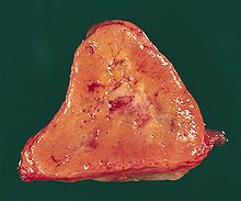 Mediastinal paraganglioma. The cut surface of a 3.9 x 3.5 x 2.5 cm tumor is triangular, with a bulging peripheral portion and a somewhat fibrotic center. It was surrounded by the heart, left lower lobe of the lung, aorta, esophagus, and diaphragm, and had been 1.8 cm in diameter 7 years before.

The paragangliomas appear grossly as sharply circumscribed polypoid masses and they have a firm to rubbery consistency. They are highly vascular tumors and may have a deep red color.
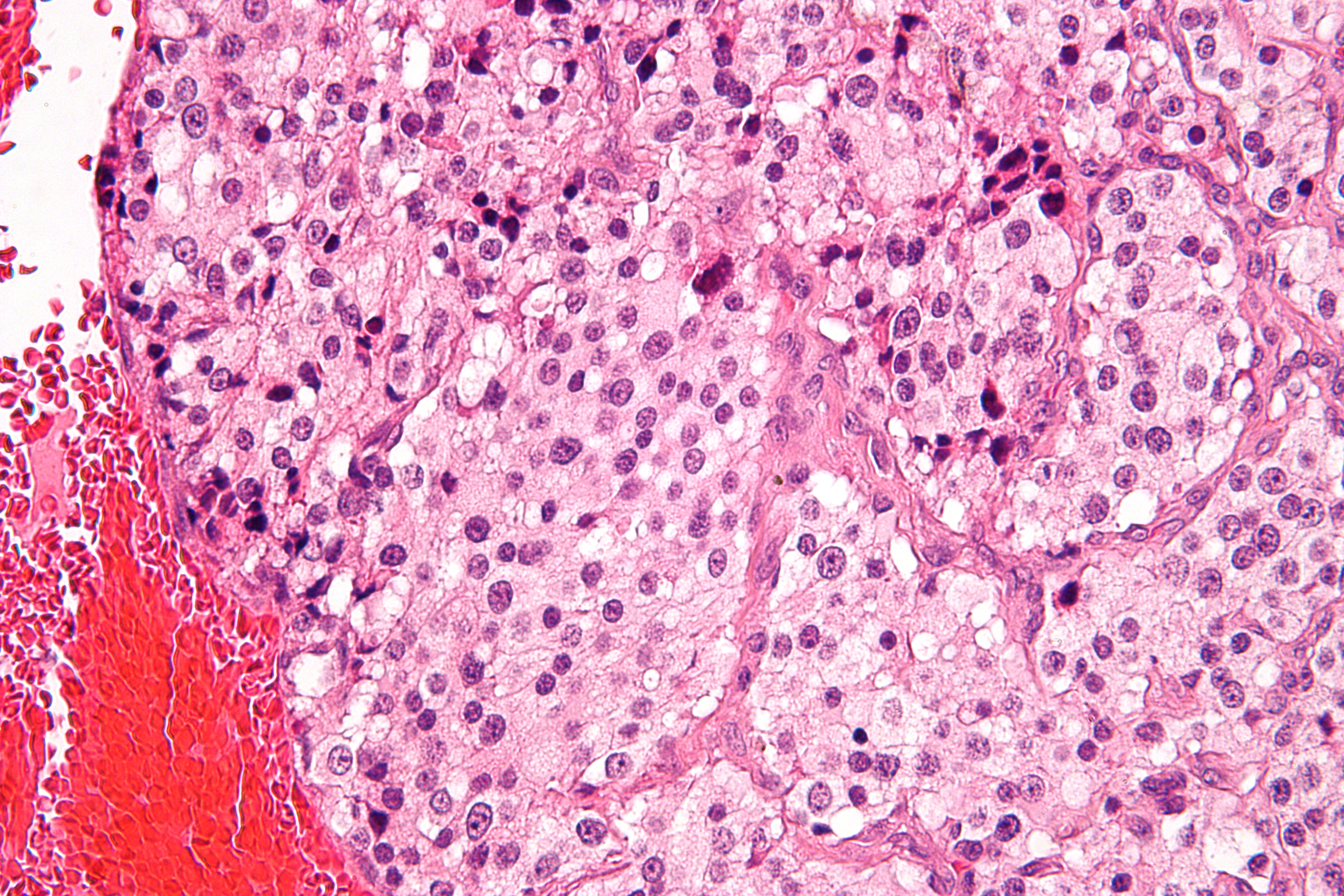
On microscopic inspection, the tumor cells are readily recognized. Individual tumor cells are polygonal to oval and are arranged in distinctive cell balls, called Zellballen.[2] These cell balls are separated by fibrovascular stroma and surrounded by sustentacular cells.
By light microscopy, the differential diagnosis includes related neuroendocrine tumors, such as carcinoid tumor, neuroendocrine carcinoma, and medullary carcinoma of the thyroid; middle ear adenoma; and meningioma.
With immunohistochemistry, the chief cells located in the cell balls are positive for chromogranin, synaptophysin, neuron specific enolase, serotonin, neurofilament and Neural cell adhesion molecule; they are S-100 protein negative. The sustentacular cells are S-100 positive and focally positive for glial fibrillary acid protein. By histochemistry, the paraganglioma cells are argyrophilic, periodic acid Schiff negative, mucicarmine negative, and argentaffin negative.
Sites of origin
About 85% of paragangliomas develop in the abdomen; only 12% develop in the chest and 3% in the head and neck region (the latter are the most likely to be symptomatic). While most are single, rare multiple cases occur (usually in a hereditary syndrome). Paragangliomas are described by their site of origin and are often given special names:-
- Carotid paraganglioma (carotid body tumor): Is the most common of the head and neck paragangliomas. It usually presents as a painless neck mass, but larger tumors may cause cranial nerve palsies, usually of the vagus nerve and hypoglossal nerve.
- Glomus tympanicum and Glomus jugulare: Both commonly present as a middle ear mass resulting in tinnitus (in 80%) and hearing loss (in 60%). The cranial nerves of the jugular foramen may be compressed, resulting swallowing difficulty, or ipsilateral weakness of the upper trapezius and sternocleiodomastoid muscles (from compression of the spinal accessory nerve). These patients present with a reddish bulge behind an intact ear drum. This condition is also known as the "Red drum". On application of pressure to the external ear canal with the help of a pneumatic ear speculum the mass could be seen to blanch. This sign is known as "Brown's sign".
- Vagal paraganglioma: These are the least common of the head and neck paragangliomas. They usually present as a painless neck mass, but may result in dysphagia and hoarseness.
- Pulmonary paraglioma: These occur in the lung and may be either single or multiple[3].
- Other sites: Rare sites of involvement are the larynx, nasal cavity, paranasal sinuses, thyroid gland, and the thoracic inlet, as well as the bladder in extremely rare cases.
Treatment
The main treatment modalities are surgery, embolization[4] and radiotherapy.[5]
Additional images
-

Micrograph of a carotid body tumor.
-

Glomus Jugulare Tumor.
-

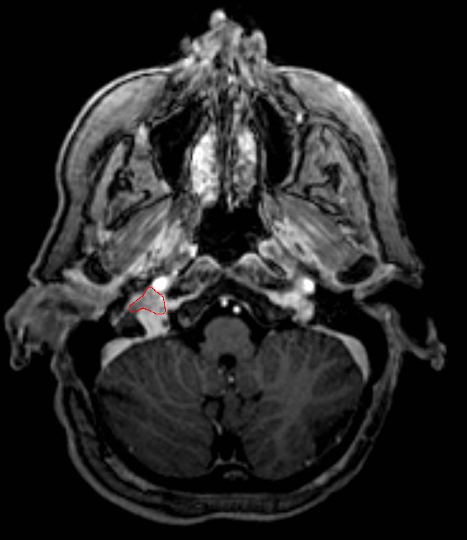
Ectopic functional paraganglioma (glomus jugulare) in a patient with VHL. T2 weighted MRI at the same location demonstrates a high signal mass consistent with a paraganglioma. Extra adrenal paragangliomas can be found in VHL (arrow).
References
- ^ Wick MR (2000). "Neuroendocrine neoplasia. Current concepts". Am. J. Clin. Pathol. 113 (3): 331–5. doi:10.1309/ETJ3-QBUK-13QD-J8FP. PMID 10705811.
- ^ Kairi-Vassilatou E, Argeitis J, Nika H, Grapsa D, Smyrniotis V, Kondi-Pafiti A (2007). "Malignant paraganglioma of the urinary bladder in a 44-year-old female: clinicopathological and immunohistochemical study of a rare entity and literature review". Eur. J. Gynaecol. Oncol. 28 (2): 149–51. PMID 17479683.
- ^ da Silva, RA; Gross JL, Haddad FJ et al. (February 2006). "Primary pulmonary paraganglioma: case report and literature review". Clinics (São Paulo) 61 (1): 83–86. doi:10.1590/S1807-59322006000100015. PMID 16532231. http://www.scielo.br/scielo.php?script=sci_arttext&pid=S1807-59322006000100015&lng=en&nrm=iso&tlng=en.
- ^ Carlsen CS, Godballe C, Krogdahl AS, Edal AL (2003). "Malignant vagal paraganglioma: report of a case treated with embolization and surgery". Auris, nasus, larynx 30 (4): 443–6. doi:10.1016/S0385-8146(03)00066-X. PMID 14656575.
- ^ Pitiakoudis M, Koukourakis M, Tsaroucha A, Manavis J, Polychronidis A, Simopoulos C (2004). "Malignant retroperitoneal paraganglioma treated with concurrent radiotherapy and chemotherapy". Clinical oncology (Royal College of Radiologists (Great Britain)) 16 (8): 580–1. PMID 15630855.
External links
- Clinical Trial information for patients with Paraganglioma
- (Description with pictures)
- [http://www.ncbi.nlm.nih.gov/bookshelf/br.fcgi?book=gene&part=paragangliomas GeneReview/NIH/UW entry on Hereditary
- Carotid Paraganglioma 3D-CT Angiogram
Paraganglioma-Pheochromocytoma Syndromes]
Gonadal tumors, paraganglioma, and glomus (ICD-O 8590-8719) Gonadal/
sex cord-gonadal stromal (8590-8679)sex cord (Granulosa cell tumour, Sertoli cell tumor)
stroma (Thecoma, Leydig cell tumor)
both (Sertoli-Leydig cell tumour, Luteoma)Paragangliomas And
Glomus tumors (8680-8719)Neuroendocrine tumor: Paraganglioma (Pheochromocytoma)
Vascular tissue neoplasm: Glomus tumor (Glomangiosarcoma)M: NEO
tsoc, mrkr
tumr, epon, para
drug (L1i/1e/V03)
Tumors: endocrine gland neoplasia (C73–C75/D34–D35, 193–194/226–227) Pancreas/
islets of LangerhansHypothalamic/
pituitary axes
+parathyroidPituitaryThyroidThyroid cancer (malignant): epithelial cell /carcinoma (Papillary, Follicular/Hurthle cell) · parafollicular cell (Medullary) · AnaplasticBenign: Thyroid adenoma · Struma ovariiParathyroidGonadssee genital neoplasiaPinealoma Pinealoblastoma · PineocytomaMEN Nervous tissue tumors/NS neoplasm/Neuroectodermal tumor (ICD-O 9350–9589) (C70–C72, D32–D33, 191–192/225) Endocrine/
sellar (9350–9379)other: PinealomaCNS
(9380–9539)Astrocytoma (Pilocytic astrocytoma, Pleomorphic xanthoastrocytoma, Fibrillary (also diffuse or lowgrade) astrocytomas, Anaplastic astrocytoma, Glioblastoma multiforme)Ependymoma · SubependymomaMultiple/unknownMature
neuronNeuroblastoma (Esthesioneuroblastoma, Ganglioneuroblastoma) · Medulloblastoma · Atypical teratoid rhabdoid tumorPrimitiveMeningiomas
(meninges)HematopoieticPNS: NST
(9540–9579)cranial and paraspinal nerves: Neurofibroma (Neurofibrosarcoma, Neurofibromatosis) · Neurilemmoma/Schwannoma (Acoustic neuroma) · Malignant peripheral nerve sheath tumornote: not all brain tumors are of nervous tissue, and not all nervous tissue tumors are in the brain (see brain metastases)
Categories:- Endocrine neoplasia


Wikimedia Foundation. 2010.