- Vestibule of the ear
-
- "Vestibulum" and "vestibule" redirect here. For other uses, see Vestibule (disambiguation).
Vestibule of the ear 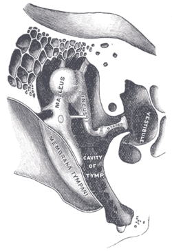
Chain of ossicles and their ligaments, seen from the front in a vertical, transverse section of the tympanum. (Vestibule visible at center right.) Latin vestibulum labyrinthi, vestibulum auris Gray's subject #232 1047 Contents
Definition
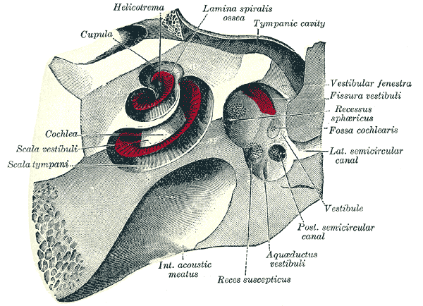
The vestibule is the central part of the osseous labyrinth, and is situated medial to the tympanic cavity, behind the cochlea, and in front of the semicircular canals.
The etymology comes from the Latin vestibulum, literally an entrance hall.
Anatomical characteristics
It is somewhat ovoid in shape, but flattened transversely; it measures about 5 mm from before backward, the same from above downward, and about 3 mm across.
In its lateral or tympanic wall is the oval window (fenestra vestibuli), closed, in the fresh state, by the base of the stapes and annular ligament.
On its medial wall, at the forepart, is a small circular depression, the recessus sphæricus, which is perforated, at its anterior and inferior part, by several minute holes (macula cribrosa media) for the passage of filaments of the acoustic nerve to the saccule; and behind this depression is an oblique ridge, the crista vestibuli, the anterior end of which is named the pyramid of the vestibule.
This ridge bifurcates below to enclose a small depression, the fossa cochlearis, which is perforated by a number of holes for the passage of filaments of the acoustic nerve which supply the vestibular end of the ductus cochlearis.
As the hinder part of the medial wall is the orifice of the aquæductus vestibuli, which extends to the posterior surface of the petrous portion of the temporal bone.
It transmits a small vein, and contains a tubular prolongation of the membranous labyrinth, the ductus endolymphaticus, which ends in a cul-de-sac between the layers of the dura mater within the cranial cavity.
On the upper wall or roof is a transversely oval depression, the recessus ellipticus, separated from the recessus sphæricus by the crista vestibuli already mentioned.
The pyramid and adjoining part of the recessus ellipticus are perforated by a number of holes (macula cribosa superior).
The apertures in the pyramid transmit the nerves to the utricle; those in the recessus ellipticus are the nerves to the ampullæ of the superior and lateral semicircular ducts.
Behind, the five orifices of the semicircular canals can be found.
There is, in the frontal view, an elliptical opening, which communicates with the scala vestibuli of the cochlea.
Additional images
-

Right osseous labyrinth (lateral view).
-

The cochlea and vestibule (view from above).
External links
See also
This article was originally based on an entry from a public domain edition of Gray's Anatomy. As such, some of the information contained within it may be outdated.

This anatomy article is a stub. You can help Wikipedia by expanding it.