- Hassall's corpuscles
-
Hassall's corpuscles 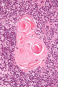
Micrograph of a thymic corpuscle. H&E stain. Gray's subject #274 1274 Hassall's corpuscles (or thymic corpuscles) are structures found in the medulla of the human thymus, formed from eosinophilic dead type VI epithelial reticular cells arranged concentrically. They are named for Arthur Hill Hassall, who discovered them in 1849.
The function of Hassall's corpuscles is currently unclear, and the absence of this structure in the murine thymus has restricted mechanistic dissection. It is known that Hassall's corpuscles are a potent source of the cytokine TSLP. In vitro, TSLP directs the maturation of dendritic cells, and increases the ability of dendritic cells to convert naive thymocytes to a Foxp3+ regulatory T cell lineage.[1][2] It is unknown if this is the physiological function of Hassall's corpuscles in vivo.
Hassall's corpuscles vary in size with diameters from 20 to more than 100μm, and tend to grow larger with age.[3]
References
- ^ * Watanabe N, Wang Y, Lee H, Ito T, Wang Y, Cao W, Liu Y (2005). "Hassall's corpuscles instruct dendritic cells to induce CD4+CD25+ regulatory T cells in human thymus.". Nature 436 (7054): 1181–5. doi:10.1038/nature03886. PMID 16121185.
- ^ "Old mystery solved, revealing origin of regulatory T cells that 'police' and protect the body"
- ^ Geneser, Finn (1999). Histologi. Munksgaard Danmark. ISBN 8762801376.
External links
- synd/1912 at Who Named It?
- UIUC Histology Subject 1247
- Histology at KUMC lymphoid-lymph03 "Hassall's Corpuscles"
- Histology at OU 61_05 - "Thymus"
- Histology at BU 07407loa - "Lymphoid Tissues and Organs: thymus, Hassall's corpuscles"
Lymphoid system (TA A13.1–2, TH H3.10, GA 8 and 9) Primary lymphoid organs Secondary lymphoid organs structural: Hilum · Trabeculae · Diaphragmatic surface of spleen · Visceral surface of spleen
Red pulp (Cords of Billroth, Marginal zone)
White pulp (Periarteriolar lymphoid sheaths, Germinal center)
blood flow: Trabecular arteries · Trabecular veinslymph flow: Afferent lymph vessels · Cortical sinuses · Medullary sinuses · Efferent lymph vessels
T cells: High endothelial venules
B cells: Primary follicle/Germinal center · Mantle zone · Marginal zone
layers: Capsule/Trabeculae · Subcapsular sinus · Cortex · Paracortex · Medulla (Medullary cord) · HilumMALT
(process mucosa)M: LMO
anat(h, u, t, a, l)/phys/depv
noco/cong/tumr
proc

This anatomy article is a stub. You can help Wikipedia by expanding it.
