- Neuromuscular junction
-
Neuromuscular junction 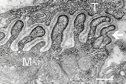
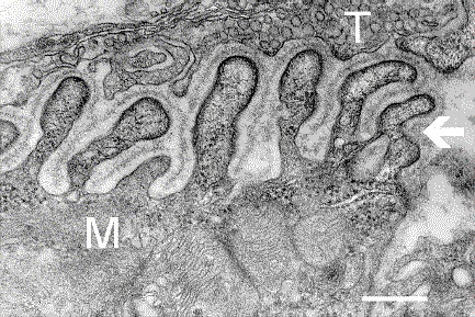
Electron micrograph showing a cross section through the neuromuscular junction. T is the axon terminal, M is the muscle fiber. The arrow shows junctional folds with basal lamina. Postsynaptic densities are visible on the tips between the folds. Scale is 0.3 µm. Source: NIMH 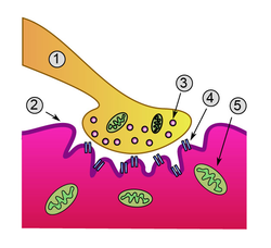
Detailed view of a neuromuscular junction:
1. Presynaptic terminal
2. Sarcolemma
3. Synaptic vesicle
4. Nicotinic acetylcholine receptor
5. MitochondrionLatin synapsis neuromuscularis; junctio neuromuscularis Code TH H2.00.06.1.02001 A neuromuscular junction (NMJ) is the synapse or junction of the axon terminal of a motor neuron with the motor end plate, the highly-excitable region of muscle fiber plasma membrane responsible for initiation of action potentials across the muscle's surface, ultimately causing the muscle to contract. In vertebrates, the signal passes through the neuromuscular junction via the neurotransmitter acetylcholine.
Contents
Mechanism of action
See also: Excitation-contraction couplingThe neuromuscular junction is the location where the neuron activates muscle to contract. This is a step in the excitation-contraction coupling of skeletal muscle.
- Upon the arrival of an action potential at the presynaptic neuron terminal, voltage-dependent calcium channels open and Ca2+ ions flow from the extracellular fluid into the presynaptic neuron's cytosol.
- This influx of Ca2+ causes neurotransmitter-containing vesicles to dock and fuse to the presynaptic neuron's cell membrane through SNARE proteins.
- Fusion of the vesicular membrane with the presynaptic cell membrane results in the emptying of the vesicle's contents (acetylcholine) into the synaptic cleft, a process known as exocytosis.
- Acetylcholine diffuses into the synaptic cleft and binds to the nicotinic acetylcholine receptors bound to the motor end plate.
- These receptors are ligand-gated ion channels, and when they bind acetylcholine, they open, allowing sodium ions to flow in and potassium ions to flow out of the muscle's cytosol.
- Because of the differences in electrochemical gradients across the plasma membrane, more sodium moves in than potassium out, producing a local depolarization of the motor end plate known as an end-plate potential (EPP).
- This depolarization spreads across the surface of the muscle fiber and continues the excitation-contraction coupling to contract the muscle.
- The action of acetylcholine is terminated when the enzyme acetylcholinesterase degrades part of the neurotransmitter (producing choline and an acetate group) and the rest of it diffuses away.
- The choline produced by the action of acetylcholinesterase is recycled — it is transported, through reuptake, back into the presynaptic terminal, where it is used to synthesize new acetylcholine molecules.[1]
Acetylcholine is a neurotransmitter synthesized in the human body from dietary choline and acetyl-CoA (ACoA). One of the first neurotransmitters discovered, the substance was originally referred to as "vagusstoff" because it was found to be released by the stimulation of the vagus nerve. Later, it was established that acetylcholine is, in fact, important in the stimulation of all muscle tissue and that its action may be either excitatory or inhibitory, depending on a number of factors. Within the body, the synaptic action of acetylcholine usually quickly comes to a halt, the neurotransmitter naturally breaking down soon after its release. However, some nerve gases are designed to thwart this breakdown, causing prolonged stimulation of the receptor cells and resulting in severe muscle spasms.
Development of the neuromuscular junction
The complex series of steps leading to the formation of the neuromuscular junction during embryonic development are only partially understood.
During development, the growing end of motor neuron axons secrete a protein known as agrin.
This protein binds to several receptors on the surface of skeletal muscle.
The receptor which seems to be required for formation of the neuromuscular junction is the MuSK protein (Muscle specific kinase).[2]
MuSK is a receptor tyrosine kinase - meaning that it induces cellular signaling by causing the release of phosphate molecules to particular tyrosines on itself, and on proteins which bind the cytoplasmic domain of the receptor.[3]
Upon activation by its ligand agrin, MuSK signals via two proteins called "Dok-7" and "rapsyn", to induce "clustering" of acetylcholine receptors (AChR).[4]
In addition to the AChR and MuSK, other proteins are then gathered, to form the endplate to the neuromuscular junction. The nerve terminates onto the endplate, forming the NMJ.
Knockout studies
These findings were demonstrated in part by mouse "knockout" studies. In mice which are deficient for either agrin or MuSK, the neuromuscular junction does not form. Further, mice deficient in Dok-7 did not form either acetylcholine receptor clusters or neuromuscular synapses.[5]
Many other proteins also comprise the NMJ, and are required to maintain its integrity.[6]
Neuromuscular block
Further information: Neuromuscular junction disease and Neuromuscular blocking drugsA block or decrease in the transmission across the neuromuscular junction can cause a complete or relative loss of muscle function. It can result from neuromuscular junction diseases or be intentionally induced with neuromuscular blocking drugs. It can also be a side effect of other drugs that are generally not classified as neuromuscular blocking drugs, such as some anesthetic drugs.[7]
The degree of neuromuscular block may be estimated by Bromage score, which originally had four grades designate with the Roman numerals I until IV, but later complemented by Breen et al with an inverse grading with Hindu-Arabic numerals:[7]
Bromage score[7] Grade Criteria Approximate
degree of blockIV 1 Complete block, inability to move feet or knees 100% III 2 Almost complete block, ability to move feet only, with inability to flex knees 66% II 3 Partial block, ability to flex knees 33% 4 Detectable weakness of hip flexion while supine, ability of full flexion of knees 5 No detectable weakness of hip flexion while supine I 6 Free movement of legs and feet, ability to perform partial knee bend 0% In unconscious patients, such as during anesthesia, neural block can be assessed by a "train-of-four" by stimulating musclesfrom surface electrodes.
See also
External links
Further reading
- Kandel, ER; Schwartz JH, Jessell TM. (2000). Principles of Neural Science (4th ed.). New York: McGraw-Hill. ISBN 0-8385-7701-6.
- Nicholls, J.G.; A.R. Martin, B.G. Wallace and P.A. Fuchs (2001). From Neuron to Brain (4th ed.). Sunderland, MA.: Sinauer Associates. ISBN 0878934391.
- Engel, A.G. (2004). Myology (3rd ed.). New York: McGraw Hill Professional. ISBN 0-07-137180-X.
References
- ^ Dale Purves, George J. Augustine, David Fitzpatrick, William C. Hall, Anthony-Samuel LaMantia, James O. McNamara, and Leonard E. White (2008). Neuroscience. 4th ed.. Sinauer Associates. pp. 121–2. ISBN 978-0-87893-697-7.
- ^ DeChiara T, Bowen D, Valenzuela D, Simmons M, Poueymirou W, Thomas S, Kinetz E, Compton D, Rojas E, Park J, Smith C, DiStefano P, Glass D, Burden S, Yancopoulos G (1996). "The receptor tyrosine kinase MuSK is required for neuromuscular junction formation in vivo". Cell 85 (4): 501–12. doi:10.1016/S0092-8674(00)81251-9. PMID 8653786.
- ^ Valenzuela D, Stitt T, DiStefano P, Rojas E, Mattsson K, Compton D, Nuñez L, Park J, Stark J, Gies D (1995). "Receptor tyrosine sinase specific for the skeletal muscle lineage: expression in embryonic muscle, at the neuromuscular junction, and after injury". Neuron 15 (3): 573–84. doi:10.1016/0896-6273(95)90146-9. PMID 7546737.
- ^ Glass D, Bowen D, Stitt T, Radziejewski C, Bruno J, Ryan T, Gies D, Shah S, Mattsson K, Burden S, DiStefano P, Valenzuela D, DeChiara T, Yancopoulos G (1996). "Agrin acts via a MuSK receptor complex". Cell 85 (4): 513–23. doi:10.1016/S0092-8674(00)81252-0. PMID 8653787.
- ^ Okada K, Inoue A, Okada M, Murata Y, Kakuta S, Jigami T, Kubo S, Shiraishi H, Eguchi K, Motomura M, Akiyama T, Iwakura Y, Higuchi O, Yamanashi Y (2006). "The muscle protein Dok-7 is essential for neuromuscular synaptogenesis". Science 312 (5781): 1802–5. doi:10.1126/science.1127142. PMID 16794080. link
- ^ Strochlic L, Cartaud A, Cartaud J (2005). "The synaptic muscle-specific kinase (MuSK) complex: new partners, new functions". Bioessays 27 (11): 1129–35. doi:10.1002/bies.20305. PMID 16237673.
- ^ a b c AnaesthesiaUK > Bromage scale Created: 30/11/2004
Histology: muscle tissue (TH H2.00.05, H3.3) Smooth
muscleStriated
muscleCostamere/
DAPCMembrane/
extracellularIntracellularDystrophin · Dystrobrevin (A, B) · Syntrophin (A, B1, B2, G1, G2) · Syncoilin · Dysbindin · Synemin/desmuslin
related: NOS1 · Caveolin 3GeneralNeuromuscular junction · Motor unit · Muscle spindle · Excitation-contraction coupling · Sliding filament mechanismBothFiberCellsOtherOther/
ungroupedCategories:- Somatic motor system
- Skeletal muscle
- Neurophysiology

Wikimedia Foundation. 2010.