- Thymus
Infobox Anatomy
Name = PAGENAME
Latin =
GraySubject = 274
GrayPage = 1273
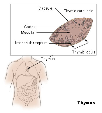
Caption = Thymus

Caption2 = The thymus of a full-term fetus, exposed in situ.
Precursor = thirdbranchial pouch
System =
Artery = derived frominternal mammary artery ,superior thyroid artery , andinferior thyroid artery
Vein =
Nerve =vagus
Lymph = tracheobronchial , parasternal
MeshName = Thymus+gland
MeshNumber = A10.549.750
DorlandsPre = t_10
DorlandsSuf = 12807749In
human anatomy , the thymus is an organ located in the upperanterior portion of the chest cavity just behind thesternum . Hormones produced by this organ stimulate the production of certain infection-fighting cells. It is of central importance in the maturation ofT cells .History
The thymus was known to the Ancient Greeks.
Galen was the first to note that the size of the organ changed over the duration of a person's life. [cite journal |author=Nishino M, Ashiku SK, Kocher ON, Thurer RL, Boiselle PM, Hatabu H |title=The thymus: a comprehensive review |journal=Radiographics |volume=26 |issue=2 |pages=335–48 |year=2006 |pmid=16549602 |doi=10.1148/rg.262045213|url=http://radiographics.rsnajnls.org/cgi/content/full/26/2/335]Due to the large numbers of
apoptotic lymphocytes, the thymus was originally dismissed as a "lymphocyte graveyard", without functional importance. The importance of the thymus in theimmune system was discovered in 1961 byJacques Miller , by surgically removing the thymus from three day old mice, and observing the subsequent deficiency in a lymphocyte population, subsequently named T cells after the organ of their origin. [cite journal |author=Miller JF |title=The discovery of thymus function and of thymus-derived lymphocytes |journal=Immunol. Rev. |volume=185 |issue= |pages=7–14 |year=2002 |pmid=12190917| doi = 10.1034/j.1600-065X.2002.18502.x ] [cite journal |author=Miller JF |title=Events that led to the discovery of T-cell development and function--a personal recollection |journal=Tissue Antigens |volume=63 |issue=6 |pages=509–17 |year=2004 |pmid=15140026 |doi=10.1111/j.0001-2815.2004.00255.x] Recently advances inimmunology have allowed the fine dissection of the function of the thymus in T cell maturation.Function
In the two thymic lobes,
lymphocyte precursors from the bone-marrow becomethymocytes , and subsequently mature into T cells. Once mature, T cells emigrate from the thymus and constitute the peripheral T cell repertoire responsible for directing many facets of theadaptive immune system . Loss of the thymus at an early age through genetic mutation (as in DiGeorge Syndrome [cite web |url=http://www.emedicine.com/med/topic567.htm |title=DiGeorge Syndrome |accessdate=2008-09-29 |author=Hussain, I., P.H. Win and S. Guduri |publisher=eMedicine |date=February 2, 2006 ] ) or surgical removal results in severe immunodeficiency and a high susceptibility to infection. [Miller JF. The discovery of thymus function and of thymus-derived lymphocytes. "Immunol Rev" 185:7-14, 2002. [http://www.blackwell-synergy.com/doi/abs/10.1034/j.1600-065X.2002.18502.x full text] ]The stock of T-lymphocytes is built up in early life, so the function of the thymus is diminished in adults. It is, therefore, largely degenerated in elderly adults and is barely identifiable, consisting mostly of fatty tissue. [cite web |url=http://www.tiscali.co.uk/reference/encyclopaedia/hutchinson/m0008212.html |title=Thymus |accessdate=2007-12-03]
The ability of T cells to recognize foreign antigens is mediated by the
T cell receptor . TheT cell receptor undergoes genetic rearrangement duringthymocyte maturation, resulting in each T cell bearing a unique T cell receptor, specific to a limited set ofpeptide :MHC combinations. The random nature of the genetic rearrangement results in a requirement ofcentral tolerance mechanisms to remove or inactivate those T cells which bear aT cell receptor with the ability to recognise self-peptides.Phases of thymocyte maturation
The generation of T cells expressing distinct T cell receptors occurs within the thymus, and can be conceptually divided into three phases:
# A rare population of hematopoietic progenitor cells enter the thymus from the blood, and expands by cell division to generate a large population of immature
thymocytes . [Schwarz BA, Bhandoola A. Trafficking from the bone marrow to the thymus: a prerequisite for thymopoiesis. "Immunol Rev" 209:47, 2006. [http://www.blackwell-synergy.com/doi/abs/10.1111/j.0105-2896.2006.00350.x full text] ]
# Immature thymocytes each make distinct T cell receptors by a process of gene rearrangement. This process is error-prone, and some thymocytes fail to make functional T cell receptors, whereas other thymocytes make T cell receptors that are autoreactive. [Sleckman BP, Lymphocyte antigen receptor gene assembly: multiple layers of regulation. "Immunol Res" 32:153-8, 2005. [http://journals.humanapress.com/index.php?option=com_opbookdetails&task=articledetails&category=humanajournals&article_code=IR:32:1:253 full text] ]Growth factors includethymopoietin andthymosin .
# Immature thymocytes undergo a process of selection, based on the specificity of their T cell receptors. This involves selection of T cells that are "functional (positive selection)", and elimination of T cells that are "autoreactive (negative selection)".Cells that pass both levels of selection are released into the
bloodstream to perform vital immune functions.Anatomy
The thymus is of a pinkish-gray color, soft, and lobulated on its surfaces. At birth it is about 5 cm in length, 4 cm in breadth, and about 6 mm in thickness.cite book |title=Anatomy of the Human Body |last=Gray |first=H. |year=1918 |chapter=4c. The Thymus |publisher=Lea & Febiger |location=Philadelphia |isbn= |pages= |url=http://www.bartleby.com/107/274.html (bartleby.com) ] The organ enlarges during childhood, and atrophies at puberty.
The thymus will, if examined when its growth is most active, be found to consist of two lateral lobes placed in close contact along the middle line, situated partly in the
thorax , partly in theneck , and extending from the fourthcostal cartilage upward, as high as the lower border of thethyroid gland . It is covered by thesternum , and by the origins of thesternohyoidei andsternothyreoidei . Below, it rests upon thepericardium , being separated from theaortic arch and great vessels by a layer offascia . In theneck , it lies on the front and sides of the trachea, behind the sternohyoidei and sternothyreoidei. The two lobes differ in size and may be united or separated.Development
Embryology
The two main components of the thymus, the lymphoid thymocytes and the thymic epithelial cells, have distinct developmental origins. The thymic epithelium is the first to develop, and appears in the form of two flask-shape endodermal diverticula, which arise, one on either side, from the third
branchial pouch (pharyngeal pouch), and extend lateralward and backward into the surrounding mesoderm andneural crest -derivedmesenchyme in front of the ventralaorta .Here they meet and become joined to one another by connective tissue, but there is never any fusion of the thymus tissue proper. The pharyngeal opening of each diverticulum is soon obliterated, but the neck of the flask persists for some time as a cellular cord. By further proliferation of the cells lining the flask, buds of cells are formed, which become surrounded and isolated by the invading mesoderm. Additional portions of thymus tissue are sometimes developed from the fourth
branchial pouches . [EmbryologySwiss|qblood/lymphat03]During the late stages of the development of the thymic epithelium,
hematopoietic lymphoid cells from bone-marrow precursors immigrate into the thymus and are aggregated to form lymphoid follicles.The thymus continues to grow between birth and puberty and then begins to
atrophy , a process directed by the high levels of circulating sex hormones. Proportional to thymic size, thymic activity (T cell output) is most active beforepuberty . Upon atrophy, the size and activity are dramatically reduced, and the organ is primarily replaced withfat (a phenomenon known as "organ involution "). The atrophy is due to the increased circulating level ofsex hormones , and chemical or physical castration of an adult results in the thymus increasing in size and activity. [Sutherland JS. Activation of thymic regeneration in mice and humans following androgen blockade. "J Immunol" 2005 15;175(4):2741-53] Patients with theautoimmune disease Myasthenia gravis commonly (70%) are found to have thymichyperplasia or malignancy. [cite book
last = Kumar
first = Parveen, Michael Clark
title = Clinical Medicine 5th edn.
publisher =Saunders
date = 2002
pages = 1222
isbn = 0-702-02606-9 ] The reason or order of these cirumstances has yet to be determined by medical scientists.tructure
Each lateral lobe is composed of numerous
lobule s held together by delicateareolar tissue ; the entire organ being enclosed in an investing capsule [BUHistology|07403loa] of a similar but denser structure. The primary lobules vary in size from that of a pin's head to that of a small pea, and are made up of a number of smallnodule s or follicles.The follicles are irregular in shape and are more or less fused together, especially toward the interior of the organ. Each follicle is from 1 to 2 mm in diameter and consists of a
medullary and a cortical portion [BUHistology|07401loa] , and these differ in many essential particulars from each other.Cortex
The cortical portion is mainly composed of
lymphoid cells, supported by a network of finely-branchedepithelial reticular cells , which is continuous with a similar network in the medullary portion. This network forms anadventitia to the blood vessels.The cortex is the location of the earliest events in
thymocyte development, whereT cell receptor gene rearrangement and positive selection takes place.Medulla
In the medullary portion, the
reticulum is coarser than in the cortex, the lymphoid cells are relatively fewer in number, and there are found peculiar nest-like bodies, the concentriccorpuscles of Hassall . These concentriccorpuscle s are composed of a central mass, consisting of one or moregranular cells , and of a capsule formed ofepithelioid cells. They are the remains of theepithelial tubes , which grow out from the thirdbranchial pouch es of the embryo to form the thymus. Each follicle is surrounded by a vascularplexus , from which vessels pass into the interior, and radiate from the periphery toward the center, forming a second zone just within the margin of the medullary portion. In the center of the medullary portion there are very few vessels, and they are of minute size.The medulla is the location of the latter events in thymocyte development. Thymocytes that reach the medulla have already successfully undergone
T cell receptor gene rearrangement and positive selection, and have been exposed to a limited degree of negative selection. The medulla is specialised to allow thymocytes to undergo additional rounds of negative selection to remove auto-reactive T cells from the mature repertoire. The gene AIRE is expressed in the medulla, and drives the transcription of organ-specific genes such as insulin to allow maturing thymocytes to be exposed to a more complex set of self-antigens than is present in the cortex.Vasculature
The
arteries supplying the thymus are derived from theinternal mammary , and from thesuperior thyroid andinferior thyroid s.The
veins end in the leftinnominate vein , and in thethyroid veins .The
nerves are exceedingly minute; they are derived from the vagi andsympathetic nervous system . Branches from the descendens hypoglossi andphrenic reach the investing capsule, but do not penetrate into the substance of the organ.Cancer
Two primary forms of
tumour s originate in the thymus.Tumours originating from the thymic epithelial cells are called
thymomas , and are found in about 10-15% of patients withmyasthenia gravis . Symptoms are sometimes confused withbronchitis or a strong cough because the tumor presses on the recurrent laryngeal nerve. All thymomas are potentially cancerous, but they can vary a great deal. Some grow very slowly. Others grow rapidly and can spread to surrounding tissues. Treatment of thymomas often requires surgery to remove the entire thymus. Tumours originating from the thymocytes are called thymiclymphomas .cite book |last=Huete-Garin |first=A. |coauthors=S.S. Sagel |editor=J.K.T. Lee, S.S. Sagel, R.J. Stanley and J.P. Heiken |title=Computed Body Tomography with MRI Correlation |year=2005 |publisher=Lippincott Williams & Wilkins |location=Philadelphia |isbn=0781745268 |pages=pp. 311-324 |chapter=Chapter 6: "Mediastinum", Thymic Neoplasm ]Other animals and second thymus
The thymus is also present in most vertebrates, with similar structure and function as the human thymus. Some animals have multiple secondary (smaller) thymi in the neck; this phenomenon has been reported for
mice [Terszowski G "et al." (2006) Evidence for a Functional Second Thymus in Mice. "Science". 2 March 2006. PMID 16513945] and also occurs in 5 out of 6 human fetuses. [ [http://www.nature.com/news/2006/060227/full/060227-9.html Surprise organ discovered in mice] , "Nature News", 2 March 2006] As in humans, theGuinea pig 's thymus naturally atrophies as the animal reaches adulthood, but in thehairless "Skinny pig " breed (which arose from a spontaneous laboratory mutation) it often possesses no thymic tissue whatsoever, and the organ cavity is replaced withcyst ic spaces.When animal thymic tissue is sold in a butcher shop or at a meat counter, thymus is known as
sweetbread .References
Millington GWM, Buckingham JC. (1992) Thymic peptides and neuroendocrine immune communication. J Endocrinol 133: 163-168
External links
*
=Additional
Wikimedia Foundation. 2010.