- Medial umbilical ligament
-
Medial umbilical ligament 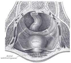
The peritoneum of the male pelvis. (Medial umbilical ligament labeled at bottom left.) 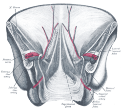
Posterior view of the anterior abdominal wall in its lower half. The peritoneum is in place, and the various cords are shining through. Latin chorda arteria umbilicalis; ligamentum umbilicale mediale Gray's subject #252 1213 The medial umbilical ligament (or cord of umbilical artery) is a paired structure found in human anatomy. It is on the deep surface of the anterior abdominal wall, and is covered by the medial umbilical folds (plica umbilicalis mediana).
Contents
Origins
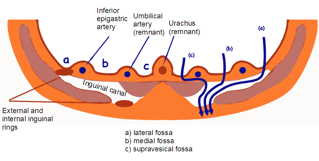
It represents the remnant of the fetal umbilical arteries, which serves no purpose in humans after birth, except for the part that becomes the adult umbilical artery.
Functions
It may be used as a landmark for surgeons exploring the medial inguinal fossa during laparoscopic inguinal hernia repair. Other than this, it has no purpose in an adult and it may be cut or damaged with impunity.
Relations
The supravesical fossa, and therefore a supravesical hernia, is medial to this structure. The medial inguinal fossa, and therefore a direct inguinal hernia, is lateral to it.
See also
- median umbilical ligament (which is a different structure)
- lateral umbilical ligament
External links
- Medial umbilical ligament
- SUNY Figs 36:01-04 - "The inguinal canal and derivation of the layers of the spermatic cord."
- SUNY Anatomy Image 7323
- SUNY Anatomy Image 7577
Additional images
-

Dissection of side wall of pelvis showing sacral and pudendal plexuses.
-

Inguinal fossae
Fetal vascular remnant ligaments (GA 6) Heart Liver Umbilical Medial umbilical ligament (see also Median umbilical ligament and Lateral umbilical fold)List of arteries of torso – abdomen (TA A12.2.12–15, GA 6.598) AA ParietalAnteriorPosteriorvisceralterminal/
common iliacAnteriorvaginal branch ♀V/IVaccompanying of ischiadic nerve · crucial anastomosisPosteriorsee arteries of lower limbsCategories:- Ligament stubs
- Abdomen
- Ligaments


Wikimedia Foundation. 2010.