- Atlas (anatomy)
-
See also: Atlas (disambiguation)
Bone: Atlas (anatomy) 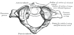
First cervical vertebra, or Atlas 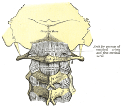
Posterior atlantoöccipital membrane and atlantoaxial ligament. (Atlas visible at center.) Gray's subject #21 99 In anatomy, the atlas (C1) is the most superior (first) cervical vertebra of the spine.
It is named for the Atlas of mythology, because it supports the globe of the head.
 Atlas
Atlas
The atlas is the topmost vertebra, and – along with the Axis – forms the joint connecting the skull and spine. The atlas and axis are specialized to allow a greater range of motion than normal vertebrae. They are responsible for the nodding and rotation movements of the head.
The atlanto-occipital joint allows the head to nod up and down on the vertebral column. The dens acts as a pivot that allows the atlas and attached head to rotate on the axis, side to side.
The Atlas' chief peculiarity is that it has no body, it is ring-like, and consists of an anterior and a posterior arch and two lateral masses.
The Atlas and Axis are important neurologically because the brain stem extends down to the Axis.
Contents
Anterior arch
The anterior arch forms about one-fifth of the ring: its anterior surface is convex, and presents at its center the anterior tubercle for the attachment of the Longus colli muscles and the anterior longitudinal ligament; posteriorly it is concave, and marked by a smooth, oval or circular facet (fovea dentis), for articulation with the odontoid process (dens) of the axis.
The upper and lower borders respectively give attachment to the anterior atlantooccipital membrane and the anterior atlantoaxial ligament; the former connects it with the occipital bone above, and the latter with the axis below.[1]
Posterior arch
 Median sagittal section through the occipital bone and first three cervical vertebræ, showing ligamentous attachments.
The posterior arch forms about two-fifths of the circumference of the ring: it ends behind in the posterior tubercle, which is the rudiment of a spinous process and gives origin to the Recti capitis posteriores minores and the ligamentum nuchae. The diminutive size of this process prevents any interference with the movements between the atlas and the skull.
The posterior part of the arch presents above and behind a rounded edge for the attachment of the posterior atlantooccipital membrane, while immediately behind each superior articular process is a groove (sulcus arteriae vertebralis), sometimes converted into a foramen by a delicate bony spiculum which arches backward from the posterior end of the superior articular process.
This groove represents the superior vertebral notch, and serves for the transmission of the vertebral artery, which, after ascending through the foramen in the transverse process, winds around the lateral mass in a direction backward and medially; it also transmits the suboccipital nerve (first spinal nerve). In a common anatomic variant the vertebral artery passes through an arcuate foramen.
On the under surface of the posterior arch, behind the articular facets, are two shallow grooves, the inferior vertebral notches. The lower border gives attachment to the posterior atlantoaxial ligament, which connects it with the axis.
Lateral masses
The lateral masses are the most bulky and solid parts of the atlas, in order to support the weight of the head.
Each carries two articular facets, a superior and an inferior.
- The superior facets are of large size, oval, concave, and approach each other in front, but diverge behind: they are directed upward, medially, and a little backward, each forming a cup for the corresponding condyle of the occipital bone, and are admirably adapted to the nodding movements of the head. Not infrequently they are partially subdivided by indentations which encroach upon their margins.
- The inferior articular facets are circular in form, flattened or slightly convex and directed downward and medially, articulating with the axis, and permitting the rotatory movements of the head.
Vertebral foramen
Just below the medial margin of each superior facet is a small tubercle, for the attachment of the transverse atlantal ligament which stretches across the ring of the atlas and divides the vertebral foramen into two unequal parts:
- the anterior or smaller receiving the odontoid process of the axis
- the posterior transmitting the spinal cord (medulla spinalis) and its membranes
This part of the vertebral canal is of considerable size, much greater than is required for the accommodation of the spinal cord.
The transverse processes are large; they project laterally and downward from the lateral masses, and serve for the attachment of muscles which assist in rotating the head. They are long, and their anterior and posterior tubercles are fused into one mass; the foramen transversarium is directed from below, upward and backward.
Development
 The atlas ossifies from 3 centers.
The atlas is usually ossified from three centers.
Of these, one appears in each lateral mass about the seventh week of fetal life, and extends backward; at birth, these portions of bone are separated from one another behind by a narrow interval filled with cartilage.
Between the third and fourth years they unite either directly or through the medium of a separate center developed in the cartilage.
At birth, the anterior arch consists of cartilage; in this a separate center appears about the end of the first year after birth, and joins the lateral masses from the sixth to the eighth year.
The lines of union extend across the anterior portions of the superior articular facets.
Occasionally there is no separate center, the anterior arch being formed by the forward extension and ultimate junction of the two lateral masses; sometimes this arch is ossified from two centers, one on either side of the middle line.
Injuries
A break in the first vertebra is referred to as a Jefferson fracture.
The pharyngeal and retropharyngeal inflammations may cause decalcification of atlas vertebra. This may lead to loosening of attachments of transverse ligament which may eventually yield. This allows the dens of axis to move and exert pressure on spinal cord causing sudden death.[citation needed]
References
- ^ Gray's Anatomy pgs.36-37 (10/27/11)
External links
- Netter, Frank. Atlas of Human Anatomy, "High Cervical Spine: C1-C2"
- Atlas at eMedicine Dictionary
This article was originally based on an entry from a public domain edition of Gray's Anatomy. As such, some of the information contained within it may be outdated.
Bones of torso (TA A02.2,3, GA 2.96–128) Vertebra General structuresbody of vertebra, vertebral arch (pedicle, lamina, vertebral notch), foramina (vertebral, intervertebral), processes (transverse, articular/zygapophysis, spinous), spinal canalUncinate process of vertebra · Transverse foramen · Anterior tubercle · Carotid tubercle · Posterior tubercle
C1 (lateral mass, anterior arch, posterior arch), C2 (dens), C3, C4, C5, C6, C7Thoracic skeleton specific ribs (1, 2, 9, 10, 11, 12, true – 1–7, false – 8–12, floating – 11–12) · parts (Angle, Tubercle, Costal groove, Neck, Head)SternumThoracic cageSuperior thoracic aperture · Inferior thoracic aperture · Intercostal space · Costal margin · Infrasternal angleCategories:- Bones of the torso


Wikimedia Foundation. 2010.