- Tarsometatarsal articulations
-
Tarsometatarsal articulations 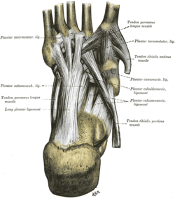
Ligaments of the sole of the foot, with the tendons of the Peronæus longus, Tibialis posterior and Tibialis anterior muscles. 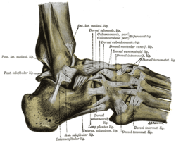
The ligaments of the foot from the lateral aspect. Latin articulationes tarsometatarsales Gray's subject #97 358 The tarsometatarsal articulations are arthrodial joints in the foot.
It is also known as the "Lisfranc joint". It is named after 18th-19th century surgeon and gynecologist, Jacques Lisfranc de St. Martin.[1]
Contents
Clinical significance
A Lisfranc fracture is common among athletes.
Anatomy
Bones
The bones entering into their formation are the first, second, and third cuneiforms, and the cuboid, which articulate with the bases of the metatarsal bones.
The first metatarsal bone articulates with the first cuneiform; the second is deeply wedged in between the first and third cuneiforms articulating by its base with the second cuneiform; the third articulates with the third cuneiform; the fourth, with the cuboid and third cuneiform; and the fifth, with the cuboid.
The bones are connected by dorsal, plantar, and interosseous ligaments.
The Dorsal Ligaments
The dorsal ligaments are strong, flat bands.
The first metatarsal is joined to the first cuneiform by a broad, thin band; the second has three, one from each cuneiform bone; the third has one from the third cuneiform; the fourth has one from the third cuneiform and one from the cuboid; and the fifth, one from the cuboid.
The Plantar Ligaments
The plantar ligaments consist of longitudinal and oblique bands, disposed with less regularity than the dorsal ligaments.
Those for the first and second metatarsals are the strongest; the second and third metatarsals are joined by oblique bands to the first cuneiform; the fourth and fifth metatarsals are connected by a few fibers to the cuboid.
The Interosseous Ligaments
The interosseous ligaments are three in number.
- The first is the strongest, and passes from the lateral surface of the first cuneiform to the adjacent angle of the second metatarsal.
- The second connects the third cuneiform with the adjacent angle of the second metatarsal.
- The third connects the lateral angle of the third cuneiform with the adjacent side of the base of the third metatarsal.
Synovial Membrane
The synovial membrane between the first cuneiform and the first metatarsal forms a distinct sac.
The synovial membrane between the second and third cuneiforms behind, and the second and third metatarsal bones in front, is part of the great tarsal synovial membrane.
Two prolongations are sent forward from it, one between the adjacent sides of the second and third, and another between those of the third and fourth metatarsal bones.
The synovial membrane between the cuboid and the fourth and fifth metatarsal bones forms a distinct sac.
From it a prolongation is sent forward between the fourth and fifth metatarsal bones.
Movements
The movements permitted between the tarsal and metatarsal bones are limited to slight gliding of the bones upon each other.
External links
References
This article was originally based on an entry from a public domain edition of Gray's Anatomy. As such, some of the information contained within it may be outdated.
Joints and ligaments of lower limbs (TA A03.6, GA 3.333) Coxal/hip femoral (iliofemoral, pubofemoral, ischiofemoral) · head of femur · transverse acetabular · acetabular labrum · capsule · zona orbicularisKnee-joint TibiofemoralCapsule · Anterior meniscofemoral ligament · Posterior meniscofemoral ligament
extracapsular: popliteal (oblique, arcuate) · collateral (medial/tibial, fibular/lateral)
intracapsular: cruciate (anterior, posterior) · menisci (medial, lateral) · transversePatellofemoralTibiofibular Superior tibiofibularInferior tibiofibularJoints of foot medial: medial of talocrural joint/deltoid (anterior tibiotalar, posterior tibiotalar, tibiocalcaneal, tibionavicular)
lateral: lateral collateral of ankle joint (anterior talofibular, posterior talofibular, calcaneofibular)Distal intertarsalOtherTarsometatarsal/LisfrancM: JNT
anat(h/c, u, t, l)/phys
noco(arth/defr/back/soft)/cong, sysi/epon, injr
proc, drug(M01C, M4)
Categories:- Musculoskeletal system stubs
- Joints


Wikimedia Foundation. 2010.