- Upper extremity of tibia
-
Bone: Upper extremity of tibia 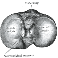
Upper surface of right tibia. (Anterior is at top.) 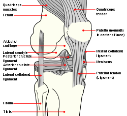
Gray's subject #61 256 The upper extremity of the tibia (or proximal extremity) is large, and expanded into two eminences, the medial condyle and lateral condyle.
Contents
Facets
The superior articular surface presents two smooth articular facets.
- The medial facet, oval in shape, is slightly concave from side to side, and from before backward.
- The lateral, nearly circular, is concave from side to side, but slightly convex from before backward, especially at its posterior part, where it is prolonged on to the posterior surface for a short distance.
The central portions of these facets articulate with the condyles of the femur, while their peripheral portions support the menisci of the knee-joint, which here intervene between the two bones.
Intercondyloid eminence
Between the articular facets, but nearer the posterior than the anterior aspect of the bone, is the intercondyloid eminence (spine of tibia), surmounted on either side by a prominent tubercle, on to the sides of which the articular facets are prolonged; in front of and behind the intercondyloid eminence are rough depressions for the attachment of the anterior and posterior cruciate ligaments and the menisci.
Surfaces
Anterior
The anterior surfaces of the condyles are continuous with one another, forming a large somewhat flattened area; this area is triangular, broad above, and perforated by large vascular foramina; narrow below where it ends in a large oblong elevation, the tuberosity of the tibia, which gives attachment to the ligamentum patellæ; a bursa intervenes between the deep surface of the ligament and the part of the bone immediately above the tuberosity.
Posterior
Posteriorly, the condyles are separated from each other by a shallow depression, the posterior intercondyloid fossa, which gives attachment to part of the posterior cruciate ligament of the knee-joint.
The medial condyle presents posteriorly a deep transverse groove, for the insertion of the tendon of the Semimembranosus.
Medial
Its medial surface is convex, rough, and prominent; it gives attachment to the tibial collateral ligament.
The lateral condyle presents posteriorly a flat articular facet, nearly circular in form, directed downward, backward, and lateralward, for articulation with the head of the fibula.
Lateral
Its lateral surface is convex, rough, and prominent in front: on it is an eminence, situated on a level with the upper border of the tuberosity and at the junction of its anterior and lateral surfaces, for the attachment of the iliotibial band.
Just below this a part of the Extensor digitorum longus takes origin and a slip from the tendon of the Biceps femoris is inserted.
Additional images
-

Right knee-joint. Anterior view.
This article was originally based on an entry from a public domain edition of Gray's Anatomy. As such, some of the information contained within it may be outdated.
Bones of lower limbs (TA A02.5.04–18, GA 2.242–277) Femur head (fovea) · neck · greater trochanter (trochanteric fossa) · lesser trochanter · intertrochanteric line · intertrochanteric crest · quadrate tubercleadductor tubercle · patellar surface · epicondyles (lateral, medial) · condyles (lateral, medial) · intercondylar fossaCrus upper extremityOtherpatella (apex of patella)Foot calcaneus (sustentaculum tali, trochlear process) · talus (body, neck, head) · navicular · cuboid · cuneiform (medial, intermediate, lateral)OtherCategories:- Bones of the lower limb


Wikimedia Foundation. 2010.