- Condylar canal
-
Bone: Condyloid canal 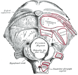
Occipital bone. Outer surface. (Condyloid canal visible at center left.) 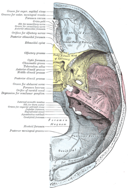
Base of the skull. Upper surface. (Condyloid canal not labeled, the occipital bone is visible at the bottom in blue, and the condyloid foramen is labeled at left, third from the bottom.) Latin canalis condylaris Gray's subject #31 131 The condylar canal (or condyloid canal) is a canal in the condyloid fossa of the lateral parts of occipital bone behind the occipital condyle. Resection of the rectus capitus posterior major and minor muscles reveals the bony recess leading to the condylar canal, which is situated posterior and lateral to the occipital condyle. It is immediately superior to the extradural vertebral artery, which is making a loop above the posterior C1 ring to enter the foramen magnum. The anteriomedial wall of the condylar canal thickens to join the foramen magnum rim and connect to the occipital condyle.
Through the condylar canal, the occipital emissary vein connects to the venous system including the suboccipital venous plexus, occipital sinus and sigmoid sinus.
It is not always present, and can have variations of being a single canal or multiple smaller canals in cluster.
Additional images
 Bilateral condylar canals (arrows) above the vertebral arteries. Dr. Victor Yang 2009
-

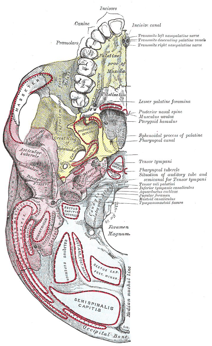
Base of skull. Inferior surface.
External links
- condylar+canal at eMedicine Dictionary
- Roche Lexicon - illustrated navigator, at Elsevier 34257.000-2
- Akram Abood Jaffar: Personal website, Anatomical variations
- Slide at uiuc.edu
This article was originally based on an entry from a public domain edition of Gray's Anatomy. As such, some of the information contained within it may be outdated.
Bones of head and neck: Foramina of the skull (and canals, fissures, meati, and hiati) (TA A02.1.00.053–097, GA 2.178–199) Anterior cranial fossa to Orbit: ethmoidal foramina (anterior, posterior)
to Nasal cavity: olfactory foramina (CN-I) · foramen cecumMiddle cranial fossa to Orbit: optic canal (CN-II) · superior orbital fissure (CN-III,IV,V1,VI)
to Pterygopalatine fossa: foramen rotundum (CN-V2) · pterygoid canal
to Infratemporal fossa: foramen ovale (CN-V3) · foramen spinosum/carotid canal
other: foramen lacerum · hiatus for greater petrosal nerve · hiatus for lesser petrosal nerve · sphenoidal emissary foramenPosterior cranial fossa internal auditory meatus/facial canal/stylomastoid foramen (CN-VII,VIII) · jugular foramen (CN-IX,X,XI) · foramen magnum (CN-XI) · hypoglossal canal (CN-XII) · condylar canal · mastoid foramenOrbit to Nasal cavity: nasolacrimal canal
to Face: supraorbital (notch, foramen) · infraorbital (foramen, groove) · zygomatic foramen (-facial, -temporal)
to Pterygopalatine fossa: inferior orbital fissure
other: Inferior orbital fissure · Fossa for lacrimal sacPterygopalatine fossa to Oral cavity: incisive canals · incisive foramen
to Nasal cavity: Foramen vomerinum · Meatus vomerinus · Fissura vomerina · Hiatus vomerinusOther Categories:- Foramina of the skull
- Musculoskeletal system stubs
-


Wikimedia Foundation. 2010.