- Cochlear nuclei
-
Brain: Cochlear nuclei 
Dissection of brain-stem. Dorsal view. ("Cochlear nucleus" is labeled on left, fifth from the bottom.) 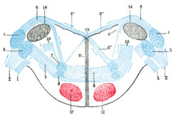
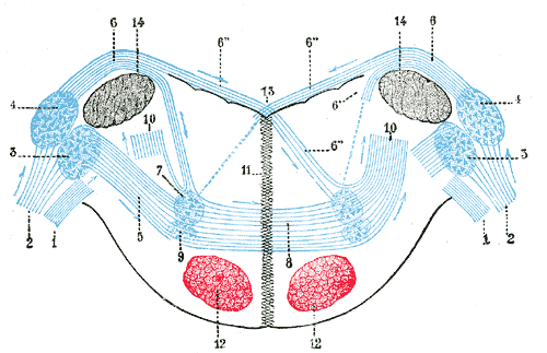
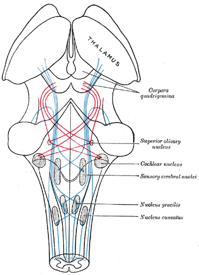
Terminal nuclei of the cochlear nerve, with their upper connections. (Schematic.) The vestibular nerve with its terminal nuclei and their efferent fibers have been suppressed. On the other hand, in order not to obscure the trapezoid body, the efferent fibers of the terminal nuclei on the right side have been resected in a considerable portion of their extent. The trapezoid body, therefore, shows only one-half of its fibers, viz., those that come from the left.
1. Vestibular nerve, divided at its entrance into the medulla oblongata.
2. Cochlear nerve.
3. Accessory nucleus of acoustic nerve.
4. Tuberculum acusticum.
5. Efferent fibers of accessory nucleus.
6. Efferent fibers of tuberculum acusticum, forming the striae medullares, with 6’, their direct bundle going to the superior olivary nucleus of the same side; 6’’, their decussating bundles going to the superior olivary nucleus of the opposite side.
7. Superior olivary nucleus.
8. Trapezoid body.
9. Trapezoid nucleus.
10. Central acoustic tract (lateral lemniscus).
11. Raphé.
12. Cerebrospinal fasciculus.
13. Fourth ventricle.
14. Inferior peduncle.Latin nuclei cochleares Gray's subject #187 788 Part of Medulla System Auditory system Artery AICA NeuroNames hier-717 The cochlear nuclei (CN) are two heterogeneous collections of neurons in the mammalian brainstem that receive input from the cochlear nerve, which carry sound information from the cochleae. The outputs from the CN are to higher regions of the auditory brainstem.
Contents
Anatomy
The CN is located at the dorso-lateral side of the brainstem, spanning the junction of the pons and medulla.
Each CN can be anatomically divided into 2 regions:
- (a) the dorsal cochlear nucleus (DCN), corresponding to the tuberculum acusticum on the dorso-lateral surface of the inferior peduncle; and
- (b) the ventral or accessory cochlear nucleus, placed between the two divisions of the nerve, on the ventral aspect of the inferior peduncle.
The ventral cochlear nucleus is further divided into the posteroventral cochlear nucleus (PVCN) and the anteroventral cochlear nucleus (AVCN).[1]
Projections to the Cochlear Nuclei
The major input to the cochlear nucleus is from the auditory nerve, a part of Cranial nerve VIII (the vestibulocochlear nerve). The auditory nerve fibers form a highly organized system of connections according to their peripheral innervation of the cochlea. Axons from the spiral ganglion cells of the lower frequencies innervate the lateral-ventral portions of the dorsal cochlear nucleus and the ventrolateral portions of the anteroventral cochlear nucleus. In contrast, the axons from the higher frequency organ of corti hair cells project to the dorsal portion of the anteroventral cochlear nucleus and the dorsal-medial portions of the dorsal cochlear nucleus. The mid frequency projections end up in between the two extremes; in this way the frequency spectrum is preserved. In this way, the cochlear nuclei inherit the tone based organization of the cochleae. This so-called tonotopic organization is preserved because only a few inner hair cells synapse on the dendrites of a nerve cell in the spiral ganglion, and the axon from that nerve cell synapes on only a very few dendrites in the cochlear nucleus.
The cochlear nuclei have long been thought to receive input only from the ipsilateral ear. There is evidence, however, for stimulation from the contralateral ear via the contralateral CN,[2] and also the somatosensory parts of the brain.[3]
Projections from the Cochlear Nuclei
There are three major projections from the cochlear nuclei. Through the medulla, one projection goes to the contralateral superior olivary complex (SOC) via the trapezoid body, whilst the other half shoots to the ipsilateral SOC. This projection is called the ventral acoustic stria (or, more commonly, the trapezoid body). Another projection, called the dorsal acoustic stria (DAS, also known as the stria of von Monakow), rises above the medulla into the pons where it hits the nucleus of the lateral lemniscus along with its kin, the intermediate acoustic stria (IAS, also known as the stria of Held). The IAS decussates across the medulla, before joining the ascending fibers in the contralateral lateral lemniscus. The lateral lemniscus contains cells of the nuclei of the lateral lemniscus, and in turn projects to the inferior colliculus. The inferior colliculus receives direct, monosynaptic projections from the superior olivary complex the contralateral dorsal acoustic stria, some classes of stellate neurons of the VCN, as well as from the different nuclei of the lateral lemniscus.
All of these inputs terminate in the inferior colliculus, although there are a few small projections that bypass the inferior colliculus and project to the medial geniculate, or other forebrain structures.
- Medial superior olive (MSO) via Trapezoid Body (TB) – Ipsilateral and contralateral stimulation for low frequency sounds.
- Lateral superior olive (LSO) directly and via TB – Ipsilateral stimulation for high frequency sounds.
- Medial Nucleus of Trapezoid body (MNTB) – Contralateral stimulation.
- Inferior colliculus – Contralateral stimulation.
- Periolivary nuclei (PON) – Ipsilateral and Contralateral stimulation.
- Lateral lemniscus (LL) and Lemniscal Nuclei (LN) – Ipsilateral and Contralateral Stimulation.
Cell types & Physiology
There are four types of principal cells found in the cochlear nuclei: Bushy cells, stellate cells, octopus cells, and fusiform cells.
- Bushy cells are found in the anterior ventral cochlear nucleus (AVCN). These can be further divided into spherical and globular bushy cells, depending on their appearance, and also their location. Within the AVCN there is an area of large spherical cells; caudal to this are smaller spherical cells, and globular cells. They have a few (1-4) very short dendrites with numerous small branching, which cause it to resemble a “bush”. The bushy cells are only found in the ventral portion of the AVCN itself. The bushy cells have specialized electrical properties that allow them to transmit timing information from the auditory nerve to more central areas of the auditory system. Some bushy cells can even improve the precision of the timing information. Bushy cells have responses very similar to those in the auditory nerve. The primary difference is that spontaneous activity is decreased by stimulation by adjacent frequencies, therefore leading to an even sharper tuning curve than seen in auditory nerve cells. These cells are usually innervated only by a few auditory nerve fibres, which dominate its firing pattern. These afferent nerve fibres wrap their terminal branches around the entire soma, creating a large synapse onto the bushy cells, called an "Endbulb of Held". Therefore, a single unit recording of an electrically stimulated bushy neuron characteristically produces exactly one action potential and constitutes the primary response.
- Stellate cells (aka multipolar cells), morphologically, have a radial, star-like dendritic tree, which is where they get their name. They are also called chopper cells, in reference to their ability to fire a regularly spaced train of action potentials for the duration of a tonal or noise stimulus. The chopping pattern is intrinsic to the electrical excitability of the stellate cell, and the firing rate depends on the strength of the auditory input more than on the frequency.
- Octopus cells are found in a small region of the Posterior Ventral Cochlear Nucleus (PVCN). The distinguishing features of these cells are their long, thick dendrites that typically emanate from one side of the cell body. Octopus cells produce an "Onset Response" to simple tonal stimuli. That is, they respond only at the onset of a specific frequency or frequency range at higher amplitudes. The octopus cells can fire with some of the highest temporal precision of any neuron in the brain. Electrical stimuli to the auditory nerve has been shown to evoke a graded post synaptic potential in the octopus cells. These EPSP’s are very brief. The octopus cells are thought to be important extracting timing information. It has been reported that these cells can respond to click trains at a rate of 800 Hz.
- Fusiform cells (also known as pyramidal cells) are found in the Dorsal Cochlear Nucleus (DCN). See the separate page concerning the DCN.
Neurotransmitters: There are four neurotransmitters responsible for transmission of neural impulses, namely, GABA, Norepinephrine, Glutamate, and Acetylcholine.
Function
The CN is the first relay station in the auditory system and can be characterized as a point of divergence in the representation of auditory information. Information is brought to the CN from the ipsilateral cochlea via the cochlear nerve. In general, the cells of the cochlear nuclei tend to preserve or even enhance the timing information that is provided by the each fiber of the cochlear nerve. The information is used by higher brainstem regions to achieve computational objectives (such as sound source location or improvement in signal to noise ratio). The cochlear nucleus receives input from each spiral ganglion, but also receives input from other parts of the brain, such as auditory cortex, pontine nuclei, trigeminal ganglion and nucleus, dorsal column nuclei and the second dorsal root ganglion. The inputs from these other areas of the brain probably play a role in sound localization.
In order to understand in more detail the specific functions of the cochlear nucleus it is first necessary to understand the way sound information is represented by the fibers of the auditory nerve. Briefly, there are around 30,000 auditory nerve fibres in each of the two auditory nerves. Each fiber is an axon of a spiral ganglion cell that represents a particular frequency of sound, and a particular range of loudness. Information in each nerve fibre is represented by the rate of action potentials as well as the particular timing of individual action potentials. The particular physiology and morphology of each cochlear nuclei cell type enhances different aspects of sound information.
See also
References
Young ED, Spirou GA, Rice JJ, Voigt HF (June 1992). "Neural organization and responses to complex stimuli in the dorsal cochlear nucleus". Philos. Trans. R. Soc. Lond., B, Biol. Sci. 336 (1278): 407–13. doi:10.1098/rstb.1992.0076. PMID 1354382. http://rstb.royalsocietypublishing.org/cgi/pmidlookup?view=long&pmid=1354382.
- ^ Middlebrooks, J.C. (2009). "Auditory System: Central Pathways". In Squire. Encyclopedia of Neuroscience. Academic Press. pp. 745–752, here: p. 745 f..
- ^ Davis KA (September 2005). "Contralateral effects and binaural interactions in dorsal cochlear nucleus". J. Assoc. Res. Otolaryngol. 6 (3): 280–96. doi:10.1007/s10162-005-0008-5. PMC 2504593. PMID 16075189. http://www.pubmedcentral.nih.gov/articlerender.fcgi?tool=pmcentrez&artid=2504593.
- ^ Shore, S.E. (2009). "Auditory/Somatosensory Interactions". In Squire. Encyclopedia of Neuroscience. Academic Press. pp. 691–5.
Additional images
-

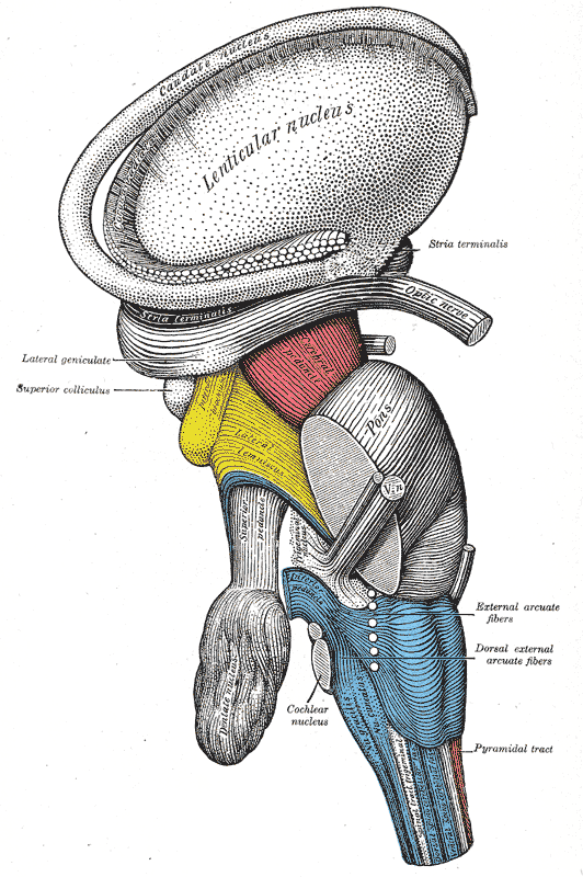
Dissection of brain-stem. Lateral view.
-
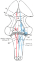
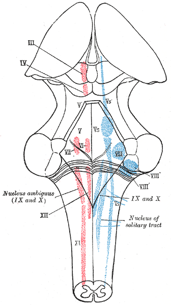
The cranial nerve nuclei schematically represented; dorsal view. Motor nuclei in red; sensory in blue.
-
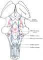
Primary terminal nuclei of the afferent (sensory) cranial nerves schematically represented; lateral view.
-
Scheme showing the course of the fibers of the lemniscus; medial lemniscus in blue, lateral in red.
-
Cross section of lower pons showing the coclear nucleus (#1) labeled at the top right.
External links
This article was originally based on an entry from a public domain edition of Gray's Anatomy. As such, some of the information contained within it may be outdated.
Human brain, rhombencephalon, metencephalon: pons (TA A14.1.05.101–604, GA 9.785) Dorsal/
(tegmentum)SurfaceWhite: Sensory/ascendingTrapezoid body/VIII · Trigeminal lemniscus (Dorsal trigeminal tract, Ventral trigeminal tract) · Medial lemniscus · Lateral lemniscus
MLF, III, IV and VI: Vestibulo-oculomotor fibers
Anterior trigeminothalamic tract · Central tegmental tractWhite: Motor/descendingICP (Vestibulocerebellar tract)
MLF, III, IV and VI: Vestibulospinal tract (Medial vestibulospinal tract, Lateral vestibulospinal tract)Other greyVentral/
(base)White: Motor/descendingSurfaceBasilar sulcusOther grey: Raphe/
reticularNerves of head and neck: the cranial nerves and nuclei (TA A14.2.01, GA 9.855) olfactory (AON->I) optic (LGN->II) oculomotor
(ON, EWN->III)trochlear (TN->IV) no significant branchestrigeminal
(PSN, TSN, MN, TMN->V)abducens (AN->VI) no significant branchesfacial (FMN, SN, SSN->VII) near origininside
facial canalvestibulocochlear
(VN, CN->VIII)glossopharyngeal
(NA, ISN, SN->IX)before jugular fossaafter jugular fossavagus
(NA, DNVN, SN->X)before jugular fossaafter jugular fossaaccessory (NA, SAN->XI) hypoglossal (HN->XII) Auditory and vestibular pathways Auditory inner ear: Hair cells → Spiral ganglion → Cochlear nerve VIII →
pons: Cochlear nuclei (Anterior, Dorsal) → Trapezoid body → Superior olivary nuclei →
midbrain: Lateral lemniscus → Inferior colliculi →
thalamus: Medial geniculate nuclei →
cerebrum: Acoustic radiation → Primary auditory cortexVestibular inner ear: Vestibular nerve VIII →
pons: Vestibular nuclei (Medial vestibular nucleus, Lateral vestibular nucleus)
cerebellum: Flocculonodular lobe
spinal cord: Vestibulospinal tract (Medial vestibulospinal tract, Lateral vestibulospinal tract)
thalamus: Ventral posterolateral nucleus
Vestibulo-oculomotor fibersM: EAR
anat(e/p)/phys/devp
noco/cong, epon
proc, drug(S2)
Categories:- Brainstem
- Neuroanatomy
- Auditory system


Wikimedia Foundation. 2010.