Cingulate cortex — Brain: Cingulate cortex Medial surface of left cerebral hemisphere … Wikipedia
Cingulate sulcus — Brain: Cingulate sulcus Medial surface of left cerebral hemisphere. MRI showing the cingulate … Wikipedia
Cingulate gyrus — Infobox Brain Name = PAGENAME Latin = gyrus cinguli GraySubject = GrayPage = Caption = Medial surface of left cerebral hemisphere. Caption2 = Human brain inferior medial view (Cingulate gyrus is #7) IsPartOf = Components = Anterior cingulate… … Wikipedia
Posterior column-medial lemniscus pathway — Originating in peripheral sensory receptors, the posterior column medial lemniscus pathway transmits fine touch and conscious proprioceptive information to the brain. Latin via column … Wikipedia
Posterior thoracic nucleus — Diagram showing a few of the connections of afferent (sensory) fibers of the posterior root with the efferent fibers from the ventral column and with the various long ascending fasciculi. (Dorsal nucleus labeled at center right.) … Wikipedia
Posterior external arcuate fibers — Diagram showing the course of the arcuate fibers. (Testut.) 1. Medulla oblongata anterior surface. 2. Anterior median fissure. 3. Fourth ventricle. 4. Inferior olivary nucleus, with the accessory olivary nuclei. 5. Gracile nucleus … Wikipedia
cingulate sulcus — sulcus cinguli [TA] a long, irregularly shaped sulcus on the medial surface of a hemisphere, which separates the cingulate gyrus below from the medial surface of the superior frontal gyrus and the paracentral lobule above. At its posterior end it … Medical dictionary
Anterior cingulate cortex — Infobox Brain Name = PAGENAME Latin = GraySubject = GrayPage = Caption = Medial surface of left cerebral hemisphere. Caption2 = IsPartOf = Components = Artery = Vein = BrainInfoType = hier BrainInfoNumber = 143 MeshName = MeshNumber = DorlandsPre … Wikipedia
nucleus lateralis posterior thalami — [TA] lateral posterior nucleus of thalamus: a nucleus in the ventral lateral part of the thalamus having major connections with the cingulate gyrus … Medical dictionary
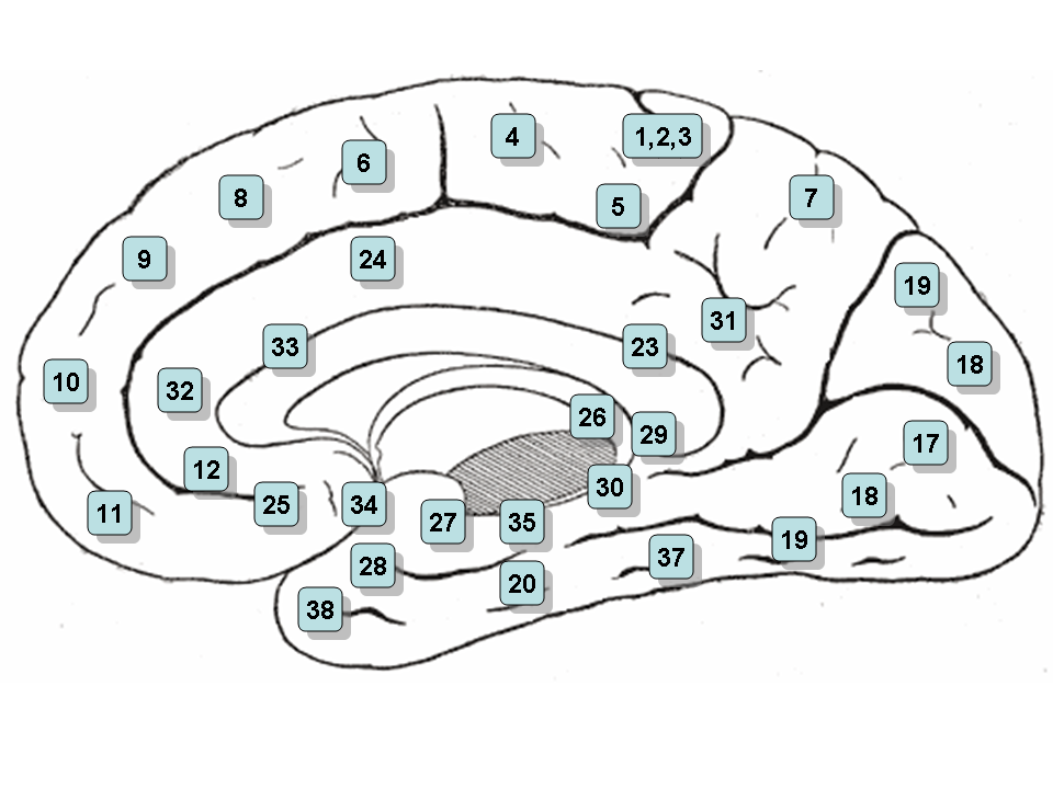
Brodmann area 23 — (BA23) is a region in the brain corresponding to some portion of the posterior cingulate cortex. It lies between Brodmann area 30 and Brodmann area 31 and is located on the medial wall of the cingulate gyrus between the callosal sulcus and the… … Wikipedia