- Medial pterygoid plate
-
Bone: Medial pterygoid plate 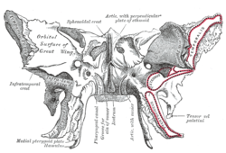
Sphenoid bone. Anterior and inferior surfaces. (Medial pterygoid plate labeled at bottom left.) 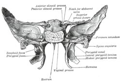
Sphenoid bone. Upper and posterior surfaces. (Medial pterygoid lamina labeled at center right.) Latin lamina medialis processus pterygoidei Gray's subject #35 151 The medial pterygoid plate (or medial pterygoid lamina) of the sphenoid is narrower and longer than the lateral pterygoid plate; it curves lateralward at its lower extremity into a hook-like process, the pterygoid hamulus, around which the tendon of the Tensor veli palatini glides.
The lateral surface of this plate forms part of the pterygoid fossa, the medial surface constitutes the lateral boundary of the choana or posterior aperture of the corresponding nasal cavity.
Superiorly the medial plate is prolonged on to the under surface of the body as a thin lamina, named the vaginal process, which articulates in front with the sphenoidal process of the palatine and behind this with the ala of the vomer.
The angular prominence between the posterior margin of the vaginal process and the medial border of the scaphoid fossa is named the pterygoid tubercle, and immediately above this is the posterior opening of the pterygoid canal.
On the under surface of the vaginal process is a furrow, which is converted into a canal by the sphenoidal process of the palatine bone, for the transmission of the pharyngeal branch of the internal maxillary artery and the pharyngeal nerve from the sphenopalatine ganglion.
The pharyngeal aponeurosis is attached to the entire length of the posterior edge of the medial plate, and the Constrictor pharyngis superior takes origin from its lower third.
Projecting backward from near the middle of the posterior edge of this plate is an angular process, the processus tubarius, which supports the pharyngeal end of the auditory tube.
The anterior margin of the plate articulates with the posterior border of the vertical part of the palatine bone.
Additional images
-

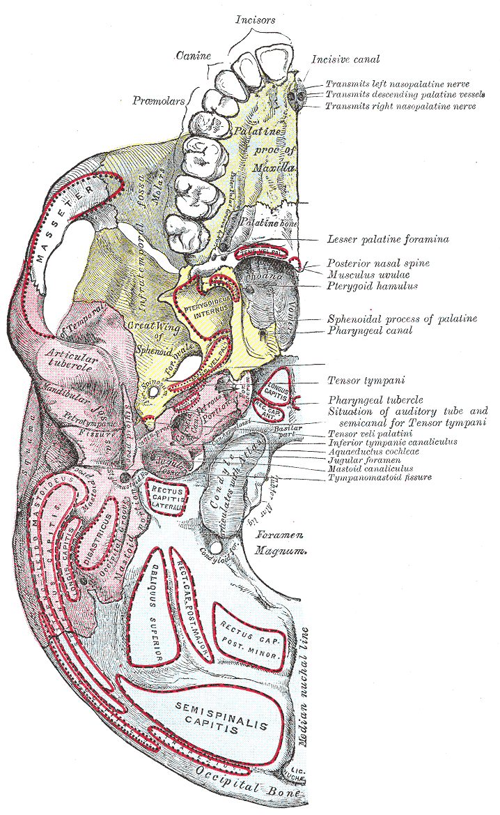
Base of skull. Inferior surface.
External links
- SUNY Figs 22:4b-05
- Roche Lexicon - illustrated navigator, at Elsevier 25420.000-1
- Roche Lexicon - illustrated navigator, at Elsevier 34257.000-1
This article was originally based on an entry from a public domain edition of Gray's Anatomy. As such, some of the information contained within it may be outdated.
Categories:- Bones of the head and neck
- Musculoskeletal system stubs
-


Wikimedia Foundation. 2010.