- Medial collateral ligament
-
Ligament: Medial collateral ligament 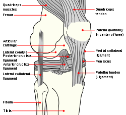
Diagram of the right knee. (Medial collateral ligament labeled at center right.) Latin ligamentum collaterale tibiale Gray's subject #93 341 From medial epicondyle of the femur To medial condyle of tibia MeSH A02.513.514.162.600 Dorlands/Elsevier l_09/12491979 The medial collateral ligament of the knee is one of the four major ligaments of the knee. It is on the medial (inner) side of the knee joint in humans and other primates. It is also known as the tibial collateral ligament, or abbreviated as the MCL.
Contents
Structure
It is a broad, flat, membranous band, situated slightly posterior on the medial side of the knee joint. It is attached proximally to the medial condyle of femur immediately below the adductor tubercle; below to the medial condyle of the tibia and medial surface of its body. It resists forces that would push the knee medially, which would otherwise produce valgus deformity.
The fibers of the posterior part of the ligament are short and incline backward as they descend; they are inserted into the tibia above the groove for the semimembranosus muscle.
The anterior part of the ligament is a flattened band, about 10 centimeters long, which inclines forward as it descends.
It is inserted into the medial surface of the body of the tibia about 2.5 centimeters below the level of the condyle.
Crossing on top of the lower part of the MCL is the pes anserinus, the joined tendons of the sartorius, gracilis, and semitendinosus muscles; a bursa is interposed between the two.
The MCL's deep surface covers the inferior medial genicular vessels and nerve and the anterior portion of the tendon of the semimembranosus muscle, with which it is connected by a few fibers; it is intimately adherent to the medial meniscus.
Embryologically and phylogenically, the ligament represents the distal portion of the tendon of adductor magnus muscle. In lower animals, adductor magnus inserts into the tibia. Because of this, the ligament occasionally contains muscle fibres. This is an atavistic variation.
Causes of injury
An MCL injury can be very painful and is caused by a valgus stress to a slightly bent knee, often when landing, bending or on high impact. Depending on the grade of the injury, the lowest grade (grade 1) can take between 2 and 10 weeks for the injury to fully heal. Recovery times for grades 2 and 3 are difficult to predict because of the amount of damage done can take weeks to several months. It is difficult to apply pressure on the injured leg for at least a few days.
Skiing
The most common knee structure damaged in skiing is the medial collateral ligament, although the carve turn has diminished the incidence somewhat.[1]
American football
MCL strains and tears are fairly common in American football. Mostly the center and the guards are ones who get this injury, due to the grip trend on their cleats(sometimes a helmet hits their knee). The number of football players who get this injury has increased in recent years. Companies are currently trying to develop better cleats that will prevent the injury.
Treatment
Treatment of a partial tear or stretch injury is usually conservative. This includes measures to control inflammation as well as bracing. Kannus has shown good clinical results with conservative care of grade II sprains, but poor results in grade III sprains.[2] As a result, more severe grade III and IV injuries to the MCL that lead to ongoing instability may require arthroscopic surgery. However, the medical literature considers surgery for most MCL injuries to be controversial.[3] Since isolated MCL injuries are uncommon, surgery is often focused on ACL replacement or repair with combined surgical approaches being common.
For higher grade tears of the MCL with ongoing instability, the MCL can be sutured or replaced. Other non-surgical approaches for more severe MCL injuries may include prolotherapy, which has been shown by Reeves in a small RCT to reduce translation on KT-1000 arthrometer versus placebo.[4] The future of non-surgical care for a non-healing MCL injury with laxity (partial ligament tear) is likely bioengineering. Fan et al. (2008) have demonstrated that knee ligament reconstruction is possible using mesenchymal stem cells and a silk scaffold.[5]
Additional images
-

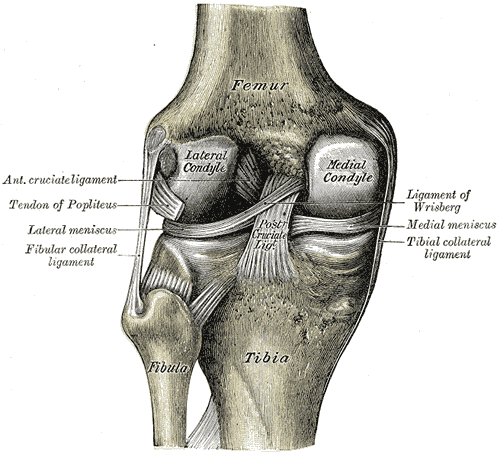
Right knee-joint. Anterior view.
-

Right knee-joint. Posterior view.
-

Left knee-joint from behind, showing interior ligaments.
-

Capsule of right knee-joint (distended). Posterior aspect.
External links
- The KNEEguru - educational site packed with knee content with sections on medial collateral ligament injuries
- SUNY Figs 17:06-04
- Medial+collateral+ligament at eMedicine Dictionary
- lljoints at The Anatomy Lesson by Wesley Norman (Georgetown University) (antkneejointopenflexed)
References
- ^ Knee injuries
- ^ Kannus P (January 1988). "Long-term results of conservatively treated medial collateral ligament injuries of the knee joint". Clinical Orthopaedics and Related Research (226): 103–12. PMID 3335084.
- ^ Indelicato PA (January 1995). "Isolated Medial Collateral Ligament Injuries in the Knee". The Journal of the American Academy of Orthopaedic Surgeons 3 (1): 9–14. PMID 10790648. http://www.jaaos.org/cgi/pmidlookup?view=long&pmid=10790648.
- ^ Reeves KD, Hassanein K (March 2000). "Randomized prospective double-blind placebo-controlled study of dextrose prolotherapy for knee osteoarthritis with or without ACL laxity". Alternative Therapies in Health and Medicine 6 (2): 68–74, 77–80. PMID 10710805.
- ^ Fan H, Liu H, Wong EJ, Toh SL, Goh JC (August 2008). "In vivo study of anterior cruciate ligament regeneration using mesenchymal stem cells and silk scaffold". Biomaterials 29 (23): 3324–37. doi:10.1016/j.biomaterials.2008.04.012. PMID 18462787.
Joints and ligaments of lower limbs (TA A03.6, GA 3.333) Coxal/hip femoral (iliofemoral, pubofemoral, ischiofemoral) · head of femur · transverse acetabular · acetabular labrum · capsule · zona orbicularisKnee-joint TibiofemoralCapsule · Anterior meniscofemoral ligament · Posterior meniscofemoral ligament
extracapsular: popliteal (oblique, arcuate) · collateral (medial/tibial, fibular/lateral)
intracapsular: cruciate (anterior, posterior) · menisci (medial, lateral) · transversePatellofemoralTibiofibular Superior tibiofibularInferior tibiofibularJoints of foot medial: medial of talocrural joint/deltoid (anterior tibiotalar, posterior tibiotalar, tibiocalcaneal, tibionavicular)
lateral: lateral collateral of ankle joint (anterior talofibular, posterior talofibular, calcaneofibular)Distal intertarsalOtherM: JNT
anat(h/c, u, t, l)/phys
noco(arth/defr/back/soft)/cong, sysi/epon, injr
proc, drug(M01C, M4)
Categories:- Ligaments of the lower limb
- Knee
-


Wikimedia Foundation. 2010.