- Medial geniculate nucleus
-
Brain: Medial geniculate nucleus 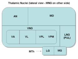
Thalamic nuclei:
MNG = Midline nuclear group
AN = Anterior nuclear group
MD = Medial dorsal nucleus
VNG = Ventral nuclear group
VA = Ventral anterior nucleus
VL = Ventral lateral nucleus
VPL = Ventral posterolateral nucleus
VPM = Ventral posteromedial nucleus
LNG = Lateral nuclear group
PUL = Pulvinar
MTh = Metathalamus
LG = Lateral geniculate nucleus
MG = Medial geniculate nucleus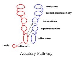
auditory pathway (Medial geniculate body labeled at upper right, second from top) Latin corpus geniculatum mediale Gray's subject #188 806 Part of Thalamus System Auditory system Artery Striata NeuroNames hier-338 NeuroLex ID birnlex_1670 The Medial Geniculate Nucleus (MGN) or Medial Geniculate Body (MGB) is part of the auditory thalamus and represents the thalamic relay between the inferior colliculus (IC) and the auditory cortex (AC). It is made up of a number of sub-nuclei that are distinguished by their neuronal morphology and density, by their afferent and efferent connections, and by the coding properties of their neurons. It is thought that the MGB influences the direction and maintenance of attention.
Contents
Divisions
The MGB has three major divisions; ventral (VMGB), dorsal (DMGB) and medial (MMGB). Whilst the VMGB is specific to auditory information processing, the DMGB and MMGB also receive information from non-auditory pathways.
aaa Inputs Outputs VMGB * Inferior colliculus
** ICC (ipsilateral)
** ICP (contralateral)
* Reticular nucleus of the thalamus (ipsilateral)
* Auditory cortexAuditory cortex
* Anterior (AAF)
* Primary (AI)
* Posterior (PAF)DMGB * IC
** Pericentral nuclei
** External nuclei
* Auditory cortex
* Other thalamic nucleiAuditory cortex MMGB * ICC (ipsilateral)
* LLN (ipsi and contra)
* Superior colliculus
* Periolivary nuclei
* Auditory cortex
** Secondary (AII)
* Reticular nucleus of thalamus
* Somatosensory and vestibular influences are also presentAuditory Cortex
* AII (ipsilateral)
* AAF (ipsilateral)
* AI (ipsilateral)
* PAF (ipsilateral)VMGB: Ventral Subnucleus of the Medial Geniculate Body
Cell types
There are two main VMGB cell types:
- Thalamocortical Relay Cells (a.k.a. Principal Neurons): The dendritic input to these cells comes from two sets of dendritic trees oriented on opposite poles of the cell. The long axis of the relay cells lie parallel to each other running superior-inferiorly with the dendritic trees of cells within the same iso-frequency band overlapping. This is similar to the dendritic organization of the ICC, but with a different orientation. The dendrites of relay cells form a synaptic nest with ascending axons from the IC and intrathalamic interneurons. In this synapse, relay cells are excited by input from the IC axons. At the same time, they are inhibited by dendritodendritic synapses from the interneurons of the VMGB. This type of synaptic nesting is characteristic of other regions in the thalamus as well.
- Intrathalamic Interneurons: These interneurons provide inhibitory (GABA) input to the relay cells at the synaptic nests. The target of their axons however, is not clear. Some interneurons appear to target relay cells, while others target other interneurons. There is also at least one type of interneuron that appears to not be involved in the synaptic nests.
Function
The VMGB is thought to be primarily responsible for relaying frequency, intensity and binaural information to the cortex. The responses in the VMGB appear to be organized in a tonotopically similar way to those in the IC. The primary difference being that the iso-freqeuncy bands are arranged such that lateral regions are most responsive to low frequencies and medial regions are responsive to high frequencies. Spatiotopic and modulotopic maps (as in the IC) however have not been well supported by mammalian studies. Both monaural (10%) and binaural cells (90%) exist in the MGB. The monaural cells are primarily responsive to sound in the contralateral hemifield. Binaural cells are typically similar to the EE or EI type found in the IC.
DMGB: Dorsal Subnucleus of the Medial Geniculate Body
Cell types
There are a large number of cell types present in the DMGB:
At least two principal cell types have been found, along with two distinct types of interneurons. Several sub-nuclei have been identified based on morphology. No frequency-specific layering has been found in the DMGB.
Function
Many types of responses are present in the DMGB that appear to vary by sub-nuclei. Generally, the responses are broadly tuned, but some cells appear to respond only to complex stimuli. Other cells are multi modal, often responding to somatosensory as well as auditory stimuli.
MMGB: Medial Subnucleus of the Medial Geniculate Body
Cell Types
Cells in the MMGB have large irregular shaped dendritic trees. There is no clear segregation based on the source of these inputs.
Function
The MMGB seems to functionally be responsible for detection of the relative intensity and duration of a sound. It shows a wide range of responses to auditory stimuli. Binaural interactions found in the MMGB include EE, EI, and IE types. Both broadly and narrowly tuned cells have been observed. A type of intensity tuning has also been observed. In this type of cell, the response actually decreases as sound intensity increases above a specific level. Almost all cells in the MMGB appear to respond for the duration of the stimulus, and have very little adaptation. Individual cells still appear to be preferentially tuned to certain frequencies, but they often have more than one CF and are broadly tuned around these CF’s. It is not clear whether there truly is one, none, or many tonotopic organizations maps present in the MMGB. Anaesthetics tend to have large effects on cells within the MMGB, making responses difficult to study. Finally, the behaviour of MMGB cells are complicated by the fact that sensory stimulation from other modalities modifies the responsiveness of many, but not all, cells in the MMGB.
See also
Additional images
-

Thalamus
-

Deep dissection of brain-stem. Lateral view.
-

Deep dissection of brain-stem. Lateral view.
-

Dissection of brain-stem. Dorsal view.
-

Hind- and mid-brains; postero-lateral view.
-

Scheme showing central connections of the optic nerves and optic tracts.
References
External links
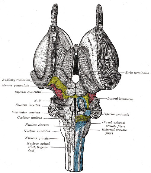
Auditory and vestibular pathways Auditory inner ear: Hair cells → Spiral ganglion → Cochlear nerve VIII →
pons: Cochlear nuclei (Anterior, Dorsal) → Trapezoid body → Superior olivary nuclei →
midbrain: Lateral lemniscus → Inferior colliculi →
thalamus: Medial geniculate nuclei →
cerebrum: Acoustic radiation → Primary auditory cortexVestibular inner ear: Vestibular nerve VIII →
pons: Vestibular nuclei (Medial vestibular nucleus, Lateral vestibular nucleus)
cerebellum: Flocculonodular lobe
spinal cord: Vestibulospinal tract (Medial vestibulospinal tract, Lateral vestibulospinal tract)
thalamus: Ventral posterolateral nucleus
Vestibulo-oculomotor fibersM: EAR
anat(e/p)/phys/devp
noco/cong, epon
proc, drug(S2)
Categories:


Wikimedia Foundation. 2010.